. 2014:2014:324713.
doi: 10.1155/2014/324713. Epub 2014 Jun 5.

Wnt pathway activation in long term remnant rat model

Affiliations

Wnt pathway activation in long term remnant rat model

E Banon-Maneus et al. Biomed Res Int. 2014.

Abstract

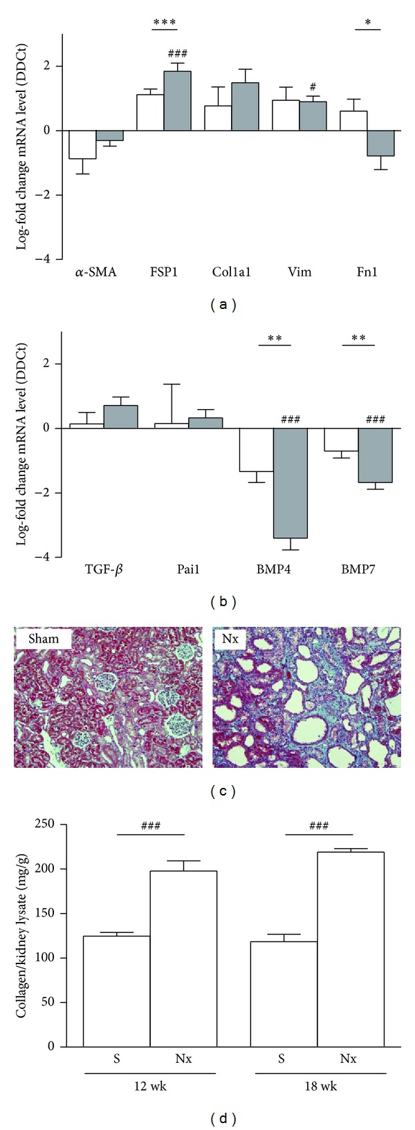
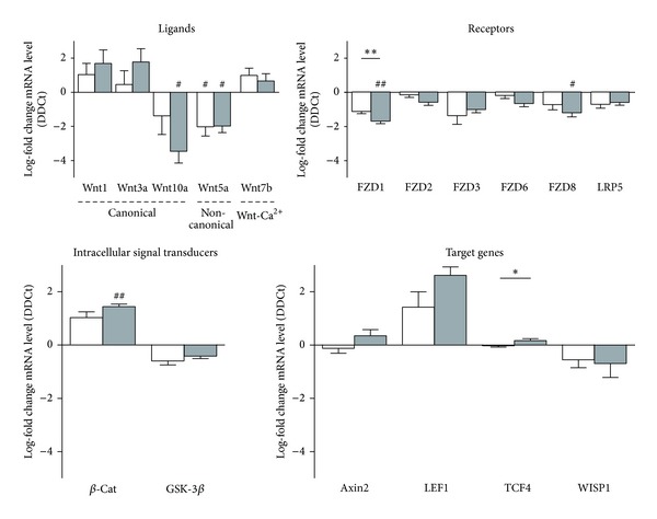
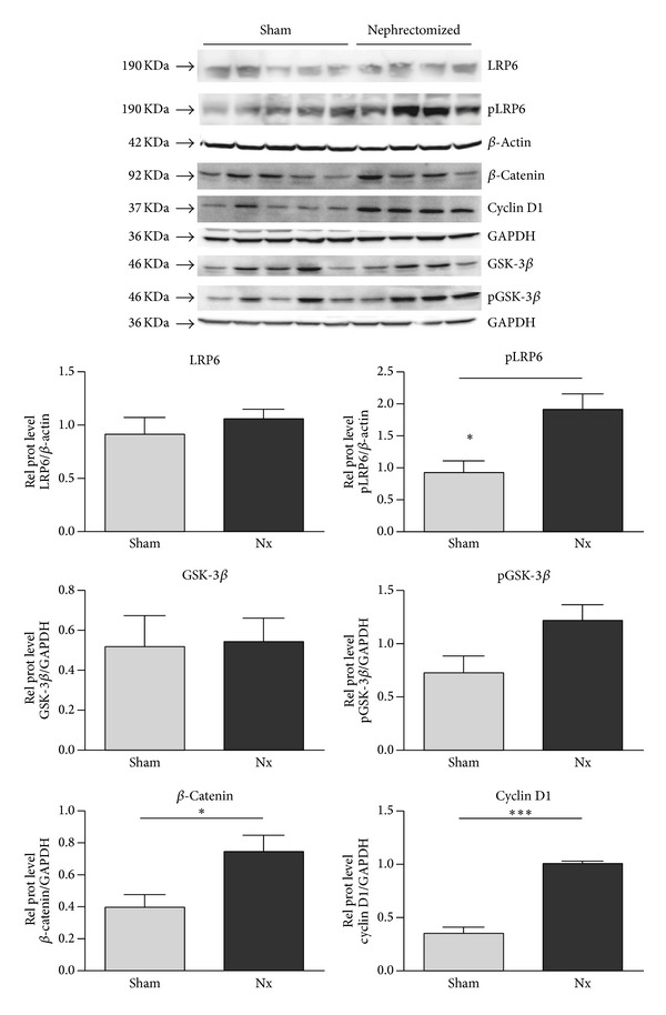
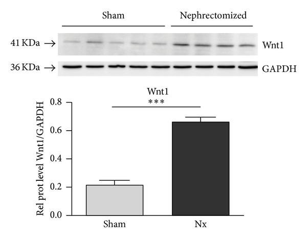
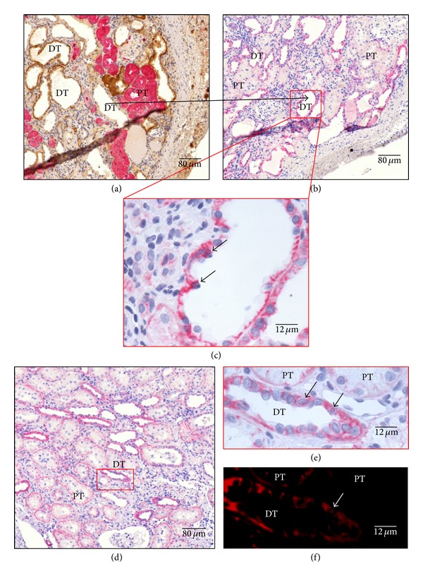
Progression of chronic kidney disease (CKD) is characterized by deposition of extracellular matrix. This is an irreversible process that leads to tubulointerstitial fibrosis and finally loss of kidney function. Wnt/ β-catenin pathway was reported to be aberrantly activated in the progressive damage associated with chronic organ failure. Extensive renal ablation is an experimental model widely used to gain insight into the mechanisms responsible for the development of CKD, but it was not evaluated for Wnt/ β-catenin pathway. This study aimed to elucidate if the rat 5/6 renal mass reduction model (RMR) is a good model for the Wnt/ β-catenin activation and possible next modulation. RMR model was evaluated at 12 and 18 weeks after the surgery, when CKD is close to end-stage kidney disease demonstrated by molecular and histological studies. Wnt pathway components were analyzed at mRNA and protein level. Our results demonstrate that Wnt pathway is active by increase of β-catenin at mRNA level and nuclear translocation in tubular epithelium as well as some target genes. These results validate the RMR model for future modulation of Wnt pathway, starting at shorter time after the surgery.


Figures

Figure 1
Figure 1
Assessment of fibrosis in rat 5/6 renal mass reduction model. (a) mRNA expression of general markers of fibroblast activation: alpha smooth muscle actin (α-SMA), fibroblast-specific protein-1 (FSP1), collagen 1a1 (Col1a1), vimentin (Vim), and fibronectin 1 (Fn1). (b) Profibrogenic mediators transforming growth factor-β (TGF-β), plasminogen activator inhibitor-1 (Pai1), and the antifibrotic bone morphogenetic proteins 4 and 7 (BMP4 and BMP7), and in rat kidneys of sham animals and 5/6 mass reduction animals by quantitative real-time PCR (qRT-PCR). Results are derived from 8-9 animals per group after 12 weeks (white columns) and 18 weeks (grey columns) after the surgery. Columns show median of log-fold change for each gene. # P > 0.05 and ### P < 0.001, two-tailed Student's t-test for unpaired observations between S and Nx groups. **P > 0.01 and ***P < 0.001, two-tailed Student's t-test for unpaired observations between 12 and 18 weeks. (c) Masson's trichrome of representative kidneys from sham or nephrectomized (Nx) rats. Representative pictures with focus on the cortex area are given. Green: collagen deposition. (d) Total collagen content was determined using the Sircol Collagen Assay. Columns are showing show median of mg collagen/g kidney lysate. ### P < 0.001, two-tailed Student's t-test for unpaired observations between S and Nx groups.
Figure 2
Figure 2
Expression of the canonical Wnt signaling components in rat 5/6 renal mass reduction model. mRNA levels of the Wnt ligands Wnt1, Wnt3a, and Wnt10a (canonical), Wnt5a (noncanonical), and Wnt7b (Ca2+ pathway); the Wnt receptors FZD1, 2, 3, 6, and 8 and LRP5; intracellular signal transducers β-catenin (β-Cat) and glucose sintase kinase3-β (GSK-3β); and the target genes axin2, lymphoid enhancer-binding factor 1 (LEF1), TCF4, and WISP1 were assessed in rat kidneys of sham animals (S) and 5/6 mass reduction animals (Nx) by quantitative real-time PCR (qRT-PCR). Results are derived from 8-9 animals per group after 12 weeks (white columns) and 18 weeks (grey columns) after the surgery. Columns show median of log-fold change for each gene. # P > 0.05 and ### P < 0.001, two-tailed Student's t-test for unpaired observations between S and Nx groups. **P > 0.01 and ***P < 0.001, two-tailed Student's t-test for unpaired observations between 12 and 18 weeks.
Figure 3
Figure 3
Activity of the canonical Wnt signal pathway in kidney homogenates of sham and nephrectomized rats. The expression of active Wnt components in kidney homogenates of sham and nephrectomized rats 18 weeks after the surgery was analyzed by immunoblotting of LRP6, phospho-LRP6, total β-catenin, cyclin D1, GSK-3β, and phospho-GSK-3β. Blotting of GDPH and β-actin served as loading controls. Results are derived from 4-5 animals per group.
Figure 4
Figure 4
Expression of Wnt1 in kidney homogenates of sham and nephrectomized rats. The expression of Wnt1 in kidney homogenates of sham and nephrectomized rats 18 weeks after the surgery was analyzed by immunoblotting. Blotting of GDPH served as loading control. Results are derived from 4-5 animals per group.
Figure 5
Figure 5
Expression and localization of β-catenin of representative kidneys in Nx rats. (a) Costain against FBPase in pink (Vulcan fast red), specific marker of proximal tubule (PT), and ATPase in brown (DAB), specific marker of distal tubule (DT), are made in serial cuts to discriminate between tubules; in (b) and (d) β-catenin stain was made in rat kidneys in pink (Vulcan fast red) and nuclei in blue (hematoxylin). Higher magnifications of tubular are shown in (c) and (e); (d) showing distal tubules with the translocated nuclei (arrows); (e) showing magnification of two proximal tubules with apical localization of β-catenin and one distal tubule with membrane localization, and short arrows indicate β-catenin translocated nucleus. As the β-catenin stain was made with Vulcan fast red, when slices were observed with a Texas Red filter clear image of translocated nuclei was obtained and false positives could be discarded; short arrow shows the confirmed 29 β-catenin translocated nucleus (f). Representative pictures with focus on the cortex area are given. Quantification of nuclei translocated β-catenin was done, related to proximal and distal epithelium surface. Stains are representative of using at least six different animals per group (magnification as indicated).

References

    1. Kim MK, Maeng YI, Sung WJ, et al. The differential expression of TGF-β1, ILK and wnt signaling inducing epithelial to mesenchymal transition in human renal fibrogenesis: an immunohistochemical study. International Journal of Clinical and Experimental Pathology. 2013;6(9):1747–1758. - PMC - PubMed
    1. von Toerne C, Schmidt C, Adams J, et al. Wnt pathway regulation in chronic renal allograft damage. American Journal of Transplantation. 2009;9(10):2223–2239. - PubMed
    1. Campistol JM, Iñigo P, Jimenez W, et al. Losartan decreases plasma levels of TGF-β1 in transplant patients with chronic allograft nephropathy. Kidney International. 1999;56(2):714–719. - PubMed
    1. Campistol JM, Iñigo P, Larios S, Bescos M, Oppenheimer F. Role of transforming growth factor-β1 in the progression of chronic allograft nephropathy. Nephrology Dialysis Transplantation. 2001;16(supplement 1):114–116. - PubMed
    1. Hwang I, Seo E-Y, Ha H. Wnt/β-catenin signaling: a novel target for therapeutic intervention of fibrotic kidney disease. Archives of Pharmacal Research. 2009;32(12):1653–1662. - PubMed

Publication types