. 2014:2014:341820.
doi: 10.1155/2014/341820. Epub 2014 Jun 4.

Multiple roles of Myd88 in the immune response to the plague F1-V vaccine and in protection against an aerosol challenge of Yersinia pestis CO92 in mice

Affiliations

Multiple roles of Myd88 in the immune response to the plague F1-V vaccine and in protection against an aerosol challenge of Yersinia pestis CO92 in mice

Jennifer L Dankmeyer et al. J Immunol Res. 2014.

Abstract

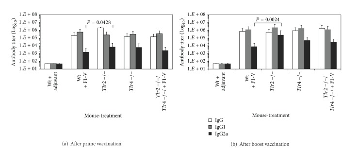
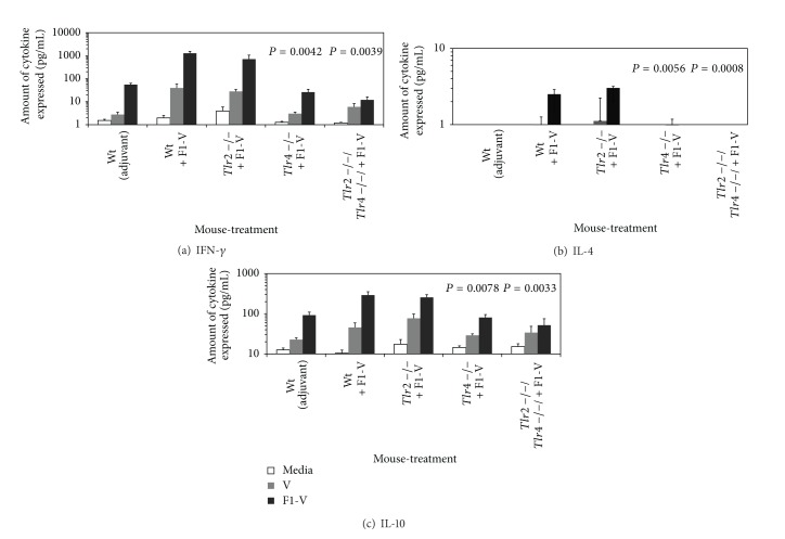
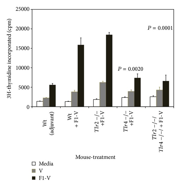
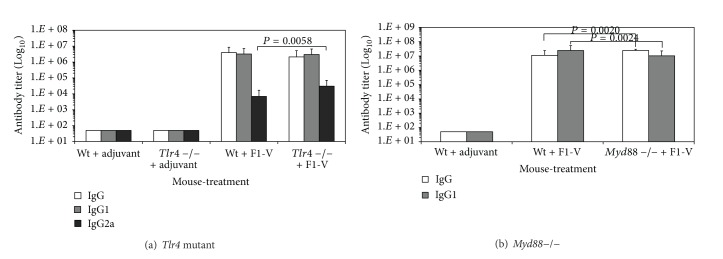
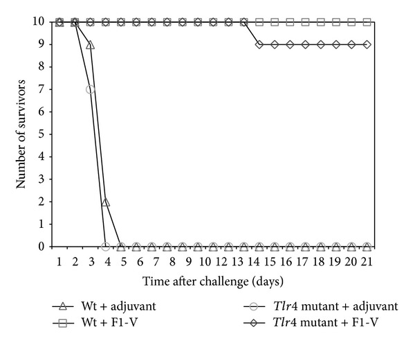
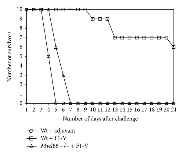
The current candidate vaccine against Yersinia pestis infection consists of two subunit proteins: the capsule protein or F1 protein and the low calcium response V protein or V-antigen. Little is known of the recognition of the vaccine by the host's innate immune system and how it affects the acquired immune response to the vaccine. Thus, we vaccinated Toll-like receptor (Tlr) 2, 4, and 2/4-double deficient, as well as signal adaptor protein Myd88-deficient mice. We found that Tlr4 and Myd88 appeared to be required for an optimal immune response to the F1-V vaccine but not Tlr2 when compared to wild-type mice. However, there was a difference between the requirement for Tlr4 and MyD88 in vaccinated animals. When F1-V vaccinated Tlr4 mutant (lipopolysaccharide tolerant) and Myd88-deficient mice were challenged by aerosol with Y. pestis CO92, all but one Tlr4 mutant mice survived the challenge, but no vaccinated Myd88-deficient mice survived the challenge. Spleens from these latter nonsurviving mice showed that Y. pestis was not cleared from the infected mice. Our results suggest that MyD88 appears to be important for both an optimal immune response to F1-V and in protection against a lethal challenge of Y. pestis CO92 in F1-V vaccinated mice.


Figures

Figure 1
Figure 1
Comparison of the class and subclass antibody titers to the Y. pestis F1-V vaccine in wild-type C3H/NeN, Tlr2, Tlr4, and Tlr2/4 deficient mice after the prime and boost vaccination. Blood was drawn 30 days after each vaccination for the antibody determination (IgG, IgG1, and IgG2a) that is reported as the geometric mean with geometric standard error of the mean. There were 6 mice in each group. There was a significant difference between the wild-type C3H/HeN mice and Tlr2 deficient C3H/HeN mice in the levels of IgG2a after the prime vaccination (P = 0.0428) and boost vaccination (P = 0.0024).
Figure 2
Figure 2
Cytokine expression by stimulated splenocytes from F1-V vaccinated wild-type C3H/HeN, Tlr2, Tlr4, and Tlr2/4 deficient mice appears to be dependent on Tlr4. Mice were given a prime-boost vaccination of F1-V (1 μg) as shown in Figure 1, and splenocytes from these same mice were prepared and stimulated for 44 h in triplicate with medium only, 4 μg of F1-V or V antigen. Culture supernatants were collected and cytokine expression was determined: (a) IFN-γ; (b) IL-4; (c) IL-10. The results are reported as mean with standard error of the mean. Statistical significance shown above the respective bar was reported for differences in cytokine expression between splenocytes from wild-type C3H/HeN and Tlr4 and Tlr2/4-deficient C3H/HeN mice stimulated with F1-V.
Figure 3
Figure 3
Proliferation by stimulated splenocytes from F1-V vaccinated wild-type C3H/HeN, Tlr2, Tlr4, and Tlr2/4-deficient mice appears to be dependent on Tlr4. Proliferation was determined after 44 h incubation in the presence of 4 μg of F1-V or V-antigen and a further 24 h incubation in the presence of 3[H]-Thymidine. Splenocytes used in the assay were prepared from the same groups of mice as shown in Figure 1. The results are reported as mean with standard error of the mean. The results on the y-axis are plotted in linear because of the overall low amount of stimulation. Statistical significance shown above the respective bar was reported for differences between the amount of proliferation between splenocytes from wild-type C3H/HeN and Tlr2 and Tlr2/4-deficient C3H/HeN mice stimulated with F1-V.
Figure 4
Figure 4
Antibody response to the plague F1-V vaccine in wild-type C57BL/6 and Myd88 deficient mice after the prime and boost vaccination. Serum for the prime vaccination was drawn 22 days after vaccination and for the boost vaccination 29 days after vaccination. All mice received 2 μg of F1-V except mice in the adjuvant only group. N for wild-type C57BL/6 mice with adjuvant and wild-type C57BL/6 mice with F1-V was 6, while for the Myd88 deficient C57BL/6 group with F1-V was 9. The titers are reported as geometric mean with geometric standard error of the mean. Significant differences in the antibody titer between the wild-type C57BL/6 mice that received F1-V and Myd88-deficient C57BL/6 mice that received F1-V which is shown above the respective bar after the prime vaccination (a).
Figure 5
Figure 5
Cytokine expression by stimulated splenocytes from F1-V vaccinated wild-type C57BL/6 or MyD88-deficient mice appears to require MyD88. Splenocytes were stimulated with F1-V (5 μg) or medium alone for approximately 45 h before collecting the supernatant and determining the amount of cytokine present: (a), IFN-γ; (b), IL-4; (c), IL-10. Cells from three different groups of mice were used (see Figure 4): (1) wild-type (Wt) C57BL/6 mice that received only adjuvant, (2) Wt C57BL/6 mice that received F1-V (2 μg), and (3) Myd88-deficient C57BL/6 mice that received F1-V (2 μg). The results are reported as mean with standard error of the mean. Statistical significance shown above the respective bar was reported for differences between the amount of cytokine expressed between the wild-type C58BL/6 and Myd88-deficient splenocytes stimulated with F1-V (P = 0.0176 and P = 0.0468) in panels (a) and (c), respectively.
Figure 6
Figure 6
Proliferation by stimulated splenocytes from F1-V vaccinated wild-type C57BL/6 mice or Myd88-deficient C57BL/6 mice requires MyD88. Proliferation of stimulated splenocytes was determined as in Figure 3 except 2 μg of F1-V was used for stimulation in 0.25 mL. The same source of splenocytes stated in Figure 5 was used. The results are reported as mean with the standard error of the mean. Statistical significance shown above the respective bars was reported for differences between the amount of proliferation between splenocytes from wild-type C57BL/6 and Myd88-deficient C57BL/6 mice stimulated with F1-V.
Figure 7
Figure 7
Antibody titers against the F1-V vaccine in (a) C3H/HeN wild-type and Tlr4 mutant C3H/HeJ mice, and (b) C57BL/6 wild-type and Myd88-deficient C57BL/6 mice vaccinated with F1-V before Y. pestis CO92 aerosol challenge. (a) There were 4 groups of mice with 10 in each group: (1) Wt+adjuvant, (2) Tlr4 mutant+adjuvant, (3) Wt+F1-V (2.9 μg), and (4) Tlr4 mutant+F1-V (2.9 μg). Antibody titers are reported from serum collected 22 days after a boost vaccination. (b) There were 3 groups of mice with 15 in groups 1 and 2 and 14 in group 3:  (1) Wt+adjuvant, (2) Wt+F1-V (2.5 μg), (3) Myd88-deficient mice+F1-V (2.5 μg). Antibody titers are reported as geometric mean with geometric standard error of the mean. Significant differences between the antibody class or subclass are reported above the respective bar between the (a) Wt+F1-V and Tlr4 mutant+F1-V or (b) Wt+F1-V and Myd88-deficient+F1-V.
Figure 8
Figure 8
Survival of F1-V vaccinated C3H/HeN wild-type and C3H/HeJ Tlr4 mutant mice after aerosol challenge with Y. pestis CO92. Mice were given two vaccinations of F1-V (2.9 μg), and 22 days after the boost vaccination, they were challenged by aerosol with 21 LD50 of Y. pestis CO92. There were four groups of mice (10 mice per group): wild-type C3H/HeN, with adjuvant only (∆); Tlr4 C3H/HeJ mutant, with adjuvant only (○); wild-type C3H/HeN, with F1-V (□); and Tlr4 C3H/HeJ mutant, with F1-V (◊). After challenge, the mice were followed for 21 days. Previous results were essentially the same when the mice in the same vaccination groups were challenged with half the dose (10 LD50) of Y. pestis CO92 except all (10/10) the F1-V vaccinated C3H/HeJ Tlr4 mutant mice survived.
Figure 9
Figure 9
Survival of F1-V vaccinated C57BL/6 wild-type and C57BL/6 Myd88-deficient mice after aerosol challenge with Y. pestis CO92. Mice were given two vaccinations of F1-V (2.5 μg) (see Figure 7(b)), and 33 days after the boost vaccination they were challenged by aerosol with 19 LD50 of Y. pestis CO92. There were three groups of mice that were challenged (10 mice per group): wild-type C57BL/6+adjuvant only (○); wild-type C57BL/6+F1-V (□); and C57BL/6 Myd88-deficient mice+ F1-V (∆). After challenge, the mice were followed for 21 days. The mean time to death (MTD) was the following: wild-type with adjuvant only: 4.50 days; wild-type with F1-V: 14.25 days; Myd88-deficient mice with F1-V: 5.90 days.
Figure 10
Figure 10
Histochemical and immunohistochemical analysis of spleens from unchallenged and challenged mice from adjuvant only C57BL/6 wild-type, F1-V vaccinated C57BL/6 wild-type, and F1-V vaccinated C57BL/6 Myd88-deficient mice. Mice were obtained from the corresponding group of mice as described in Figures 7(b) and 9. Spleens from additional mice from each group were used as controls (Unchallenged-H&E, panels (a), (d), and (g)). Regions in the spleen were labeled: white pulp, WP; red pulp, RP; and marginal zone, MZ. Spleen sections from challenged mice are shown stained (Challenged-H&E, panels (b), (e), and (h)) or probed with an anti-F1 monoclonal antibody (Challenged-Anti-F1, panels (c), (f), and (i)). The spleen section shown from Group 2, wild-type+F1-V was from a mouse that survived challenge (panels (e) and (f)). Arrows in panel (c) and (i) point to F1 positive regions in the marginal zone. Arrows in panel (f ) point to isolated F1 positive spots.

Similar articles

Cited by

References

    1. Haffkine WM. Remarks on the plague prophylactic fluid. British Medical Journal. 1897;1:1461–1462.
    1. Williamson ED, Eley SM, Griffin KF, et al. A new improved sub-unit vaccine for plague: the basis of protection. FEMS Immunology and Medical Microbiology. 1995;12(3-4):223–230. - PubMed
    1. Heath DG, Anderson GW, Welkos SL, Andrews GP, Friedlander AM, Mauro JM. A recombinant capsular F1-V antigen fusion protein vaccine protects against experimental bubonic and pneumonic plague. In: Brown F, Burton D, Doherty P, Mekalanos J, Norrby E, editors. Vaccine 97. New York, NY, USA: Cold Spring Harbor Laboratory Press; 1997. pp. 197–200.
    1. Leary SEC, Williamson ED, Griffin KF, Russell P, Eley SM, Titball RW. Active immunization with recombinant V antigen from Yersinia pestis protects mice against plague. Infection and Immunity. 1995;63(8):2854–2858. - PMC - PubMed
    1. Anderson GW, Leary SEC, Williamson ED, et al. Recombinant V antigen protects mice against pneumonic and bubonic plague caused by F1-capsule positive and -negative strains of Yersinia pestis . Infection and Immunity. 1996;64(11):4580–4585. - PMC - PubMed

Publication types

MeSH terms

LinkOut - more resources