. 2015 Mar;25(2):121-35.
doi: 10.1111/bpa.12168. Epub 2014 Sep 12.

The brainstem pathologies of Parkinson's disease and dementia with Lewy bodies

Affiliations

The brainstem pathologies of Parkinson's disease and dementia with Lewy bodies

Kay Seidel et al. Brain Pathol. 2015 Mar.

Abstract

Parkinson's disease (PD) and dementia with Lewy bodies (DLB) are among the human synucleinopathies, which show alpha-synuclein immunoreactive neuronal and/or glial aggregations and progressive neuronal loss in selected brain regions (eg, substantia nigra, ventral tegmental area, pedunculopontine nucleus). Despite several studies about brainstem pathologies in PD and DLB, there is currently no detailed information available regarding the presence of alpha-synuclein immunoreactive inclusions (i) in the cranial nerve, precerebellar, vestibular and oculomotor brainstem nuclei and (ii) in brainstem fiber tracts and oligodendroctyes. Therefore, we analyzed the inclusion pathologies in the brainstem nuclei (Lewy bodies, LB; Lewy neurites, LN; coiled bodies, CB) and fiber tracts (LN, CB) of PD and DLB patients. As reported in previous studies, LB and LN were most prevalent in the substantia nigra, ventral tegmental area, pedunculopontine and raphe nuclei, periaqueductal gray, locus coeruleus, parabrachial nuclei, reticular formation, prepositus hypoglossal, dorsal motor vagal and solitary nuclei. Additionally we were able to demonstrate LB and LN in all cranial nerve nuclei, premotor oculomotor, precerebellar and vestibular brainstem nuclei, as well as LN in all brainstem fiber tracts. CB were present in nearly all brainstem nuclei and brainstem fiber tracts containing LB and/or LN. These findings can contribute to a large variety of less well-explained PD and DLB symptoms (eg, gait and postural instability, impaired balance and postural reflexes, falls, ingestive and oculomotor dysfunctions) and point to the occurrence of disturbances of intra-axonal transport processes and transneuronal spread of the underlying pathological processes of PD and DLB along anatomical pathways.

Keywords: Parkinson's disease; alpha-synuclein; brainstem; dementia with Lewy bodies; prion-like disease.


Figures

Figure 1
Figure 1
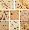
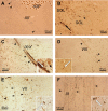
Alpha‐synuclein immunoreactive neuronal and oligodendroglial aggregation pathology in patients withP arkinson's disease ( PD ) or dementia withL ewy bodies ( DLB ). A. Alpha‐synuclein immunoreactive neuronal Lewy bodies (LB, black arrows) and Lewy neurites (LN, arrowheads) in the dorsal motor vagal nucleus (DMV) of a PD patient (case 1; Table 1). B. LN (arrowheads) and coiled body (CB, white arrow) in the vagal nerve (IX/X) of the same PD patient (case 1; Table 1). C. LB (black arrow), LN (arrowhead) and CB (white arrow) in the DMV of a PD with dementia patient (case 6; Table 1). D. LB (black arrow) and CB (white arrow) in the intermediate reticular zone (IRZ) of a Dementia with Lewy bodies (DLB) patient (case 10; Table 1). E. CB (white arrows) in the gigantocellular reticular nucleus (GI) of a PD patient (case 1; Table 1) and F. in the parvocellular reticular nucleus (PCRT) of a PD patient (case 3; Table 1). G. LB (black arrow), LN (arrowhead) and CB (white arrow) in the compact part of the pedunculopontine nucleus, compact part (PPT) of a DLB patient (case 10; Table 1). H. CB (white arrow) in the midbrain supratrochlear part of the dorsal raphe nucleus‐supratrochlear part (DR‐ST) of the same DLB patient (case 10; Table 1). (A–H: alpha‐synuclein immunostaining; 100 μm PEG sections).
Figure 2
Figure 2
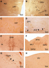
Alpha‐synuclein immunoreactive neuronal and oligodendroglial aggregation pathology in nuclei of the medulla oblongata and pontomedullary junction of patients withP arkinson's disease ( PD ) or dementia withL ewy bodies ( DLB). A. Severe alpha‐synuclein aggregation pathology in the precerebellar lateral reticular nucleus (LRT) and conspicuously affected autonomic intermediate reticular zone (IRZ) of a DLB patient (case 10; Table 1). B. Markedly involved somatomotor hypoglossal (XII) and severely affected autonomic dorsal motor vagal nuclei (DMV) of the same DLB patient. Arrowheads point to Lewy neurites (LN; case 10; Table 1). C. Marked alpha‐synuclein immunoreactive neuronal and oligodendroglial inclusions of the principal nucleus of the inferior olive (IOP) of a PD patient (case 3; Table 1). D. Alpha‐synuclein immunopositive neuronal and oligodendroglial inclusions in the spinal trigeminal nucleus (SPV) and vagal nerve (IX/X) (arrowheads) of a DLB patient (case 10; Table 1). E. Severe alpha‐synuclein immunoreactive neuronal and oligodendroglial inclusion pathologies in the gigantocellular reticular (GI) and great raphe nuclei (GRN) of the brainstem gain setting system of a DLB patient (case 11; Table 1). F. Severe alpha‐synuclein immunoreactive neuronal and oligodendroglial aggregation pathology of the oculomotor prepositus hypoglossal (PPH) and autonomic DMV, as well as markedly affected dorsal paragigantocellular reticular nucleus (DPGI) of a PD patient (case 4; Table 1). G. Prominent alpha‐synuclein immunoreactive neuronal (Lewy bodies, LB, LN) and oligodendroglial (coiled bodies, CB) aggregations in the ingestive parvocellular reticular nucleus (PCRT) of a DLB patient. Arrowheads point to LN (case 11; Table 1). H. Markedly involved medial vestibular nucleus (MV) of a PD with dementia patient (case 6; Table 1). (A–H: alpha‐synuclein immunostaining; 100 μm PEG sections). (Abbreviation: MLF = Medial longitudinal fascicle).
Figure 3
Figure 3
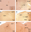
Alpha‐synuclein immunoreactive neuronal and oligodendroglial aggregation pathology in nuclei of the pons of patients with Parkinson's disease ( PD ) or dementia with Lewy bodies ( DLB ). A. Alpha‐synuclein immunoreactive Lewy bodies (LB), Lewy neurites (LN) and coiled bodies (CB) in the precerebellar pontine nuclei (PN) of a PD patient (case 1; Table 1). Inset shows alpha‐synuclein immunoreactive PN neuron. B. Alpha‐synuclein immunoreactive LB, LN and CB in the somatomotor facial nucleus (VII) and severely involved adjacent ingestive parvocellular reticular nucleus (PCRT) of a DLB patient (case 11; Table 1). C. Marked affection of the superior vestibular nucleus (SUV) of a PDD patient by the alpha‐synuclein immunoreactive aggregation pathology (case 6; Table 1). Neuronal and oligodendroglial alpha‐synuclein aggregates in D. the cell‐poor premotor oculomotor area of the excitatory burst neurons for horizontal saccades (EBR) and E. the noradrenergic locus coeruleus (LC) of a PD patient (case 3; Table 1). F. The alpha‐synuclein immunoreactive neuronal and oligodendroglial inclusion pathology marks the outlines of the precerebellar oculomotor reticulotegmental nucleus of the pons (RTTG) of a DLB patient (case 11; Table 1). (A–F alpha‐synuclein immunostaining; 100 μm PEG sections). (Abbreviations: MLF = Medial longitudinal fascicle; PNC = Pontine reticular formation, caudal nucleus; PNO = Pontine reticular formation, oral nucleus; SCP = Superior cerebellar peduncle; VII = Facial nerve).
Figure 4
Figure 4
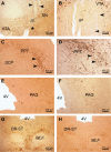
Alpha‐synuclein immunoreactive neuronal and oligodendroglial aggregation pathology in midbrain nuclei of patients withP arkinson's disease ( PD ) or dementia withL ewy bodies ( DLB ). A. Alpha‐synuclein immunoreactive neuronal (Lewy bodies, LB; Lewy neurites, LN) and oligodendroglial (coiled bodies, CB) aggregations in the dopaminergic substantia nigra (SN), nuclei of the ventral tegmental area (VTA) and oculomotor nerve (III) (arrowheads) of a Parkinson's disease with dementia (PDD) patient (case 6; Table 1). Note the substantial loss of melanin‐containing dopaminergic nerve cells and the occurrence of extraneuronal neuromelanin deposits in the SN (asterisk). B. The midbrain interpeduncular nucleus (IP) adjacent to the severely affected VTA of the same PDD patient (case 6; Table 1). Arrowheads point to LN. Alpha‐synuclein immunoreactive neuronal and oligodendroglial inclusion pathology in the cholinergic compact part of the pedunculopontine nucleus (PPT) of C. a PD patient (case 3; Table 1) and D. a DLB patient (case 10; Table 1). Arrowheads point to LN. Well‐developed alpha‐synuclein immunoreactive neuronal and oligodendroglial aggregations in the periaqueductal gray (PAG) of E. a PDD patient (case 6; Table 1) and F. a DLB patient (case 10; Table 1). Considerable amounts of LB, LN and CB in the midbrain supratrochlear part of the dorsal raphe nucleus (DR‐ST) of G. a PDD patient (case 6; Table 1) and H. a DLB patient (case 10; Table 1). (A–H: alpha‐synuclein immunostaining; 100 μm PEG sections). (Abbreviations: MLF = Medial longitudinal fascicle; SCP = Superior cerebellar peduncle; 4V = Fourth ventricle).
Figure 5
Figure 5
Alpha‐synuclein immunoreactive neuronal and oligodendroglial aggregation pathology in brainstem fiber tracts of patients with Parkinson's disease (PD) or dementia with Lewy bodies (DLB). Marked alpha‐synuclein immunoreactive neuronal (Lewy neurites, LN) and oligodendroglial (coiled bodies, CB) aggregations in A. the hypoglossal nerve (XII) of a DLB patient (arrowheads point to LN) (case 11; Table 1), B. the solitary tract (SOL) of a PD patient (arrowhead points to LN in the SOL and dark arrow points to a LB in the solitary nucleus = SOL) (case 3; Table 1), C. the vagal nerve (IX/X) of a PD patient (arrowheads point to LN and inset shows CB = white arrow) (case 6; Table 1), D. the vestibulocochlear nerve (VIII) of a PD patient (arrowheads point to LN and inset shows CB = white arrow) (case 3; Table 1), E. the facial nerve (VII) of a DLB patient (arrowheads point to LN and inset shows CB = white arrow) (case 11; Table 1), and F. the oculomotor nerve (III) of a PD with dementia patient (arrowheads point to LN and inset shows CB = white arrow) (case 6; Table 1). (A–F: alpha‐synuclein immunostaining; 100 μm PEG sections). (Abbreviations: IOP = Inferior olive, principal subnucleus).

References

    1. Ahmed Z, Asi YT, Sailer A, Lees AJ, Houlden H, Revesz T, Holton JL (2012) The neuropathology, pathophysiology and genetics of multiple system atrophy. Neuropathol Appl Neurobiol 38:4–24. - PubMed
    1. Angot E, Steiner JA, Hansen C, Li JY, Brundin P (2010) Are synucleinopathies prion‐like disorders? Lancet Neurol 9:1128–1138. - PubMed
    1. Barmack NH (2003) Central vestibular system: vestibular nuclei and posterior cerebellum. Brain Res Bull 60:511–541. - PubMed
    1. Benarroch EE (2013) Pedunculopontine nucleus: functional organization and clinical implications. Neurology 80:1148–1155. - PubMed
    1. Benarroch EE, Schmeichel AM, Sandroni P, Low PA, Parisi JE (2006) Involvement of vagal autonomic nuclei in multiple system atrophy and Lewy body disease. Neurology 66:378–383. - PubMed

Publication types

Substances