

Minimally invasive convection-enhanced delivery of biologics into dorsal root ganglia: validation in the pig model and prospective modeling in humans. Technical note
- PMID: 24995785
- PMCID: PMC5065916
- DOI: 10.3171/2014.6.JNS132364
Minimally invasive convection-enhanced delivery of biologics into dorsal root ganglia: validation in the pig model and prospective modeling in humans. Technical note
Abstract
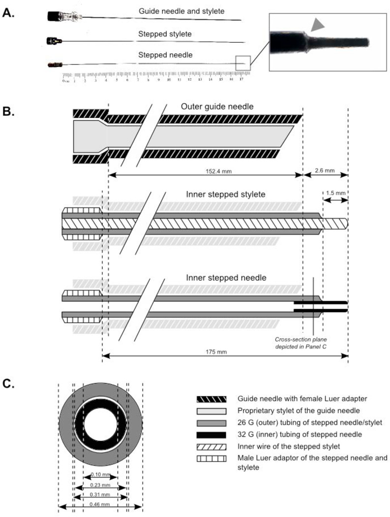
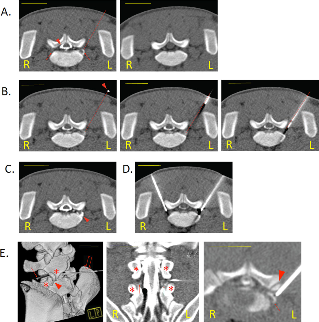
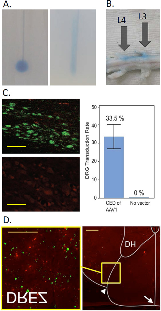
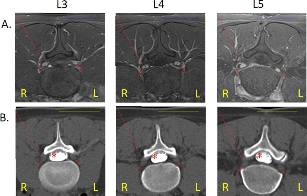
Dorsal root ganglia (DRG) are critical anatomical structures involved in nociception. Intraganglionic (IG) drug delivery is therefore an important route of administration for novel analgesic therapies. Although IG injection in large animal models is highly desirable for preclinical biodistribution and toxicology studies of new drugs, no method to deliver pharmaceutical agents into the DRG has been reported in any large species. The present study describes a minimally invasive technique of IG agent delivery in domestic swine, one of the most common large animal models. The technique utilizes CT guidance for DRG targeting and a custom-made injection assembly for convection enhanced delivery (CED) of therapeutic agents directly into DRG parenchyma. The DRG were initially visualized by CT myelography to determine the optimal access route to the DRG. The subsequent IG injection consisted of 3 steps. First, a commercially available guide needle was advanced to a position dorsolateral to the DRG, and the dural root sleeve was punctured, leaving the guide needle contiguous with, but not penetrating, the DRG. Second, the custom-made stepped stylet was inserted through the guide needle into the DRG parenchyma. Third, the stepped stylet was replaced by the custom-made stepped needle, which was used for the IG CED. Initial dye injections performed in pig cadavers confirmed the accuracy of DRG targeting under CT guidance. Intraganglionic administration of adeno-associated virus in vivo resulted in a unilateral transduction of the injected DRG, with 33.5% DRG neurons transduced. Transgene expression was also found in the dorsal root entry zones at the corresponding spinal levels. The results thereby confirm the efficacy of CED by the stepped needle and a selectivity of DRG targeting. Imaging-based modeling of the procedure in humans suggests that IG CED may be translatable to the clinical setting.
Keywords: AAV = adeno-associated virus; AAV1 = AAV serotype 1; CED = convection-enhanced delivery; CTF = CT fluoroscopy; DRG = dorsal root ganglia; EGFP = enhanced green fluorescent protein; IG = intraganglionic; PBS = phosphate-buffered saline; computed tomography; convection-enhanced delivery; dorsal root ganglia; gene therapy; intraganglionic injection; pain; peripheral nerve.
Figures
Similar articles
-
Intraneural convection enhanced delivery of AAVrh20 for targeting primary sensory neurons.Mol Cell Neurosci. 2014 May;60:72-80. doi: 10.1016/j.mcn.2014.04.004. Epub 2014 Apr 24. Mol Cell Neurosci. 2014. PMID: 24769104 Free PMC article.
-
Comparison of the efficiencies of intrathecal and intraganglionic injections in mouse dorsal root ganglion.Turk J Med Sci. 2023 Aug 11;53(5):1358-1366. doi: 10.55730/1300-0144.5702. eCollection 2023. Turk J Med Sci. 2023. PMID: 38813001 Free PMC article.
-
Rapid injection of lumbar dorsal root ganglia under direct vision: Relevant anatomy, protocol, and behaviors.Front Neurol. 2023 Apr 11;14:1138933. doi: 10.3389/fneur.2023.1138933. eCollection 2023. Front Neurol. 2023. PMID: 37114234 Free PMC article.
-
MRI guidance technology development in a large animal model for hyperlocal analgesics delivery to the epidural space and dorsal root ganglion.J Neurosci Methods. 2019 Jan 15;312:182-186. doi: 10.1016/j.jneumeth.2018.11.024. Epub 2018 Dec 1. J Neurosci Methods. 2019. PMID: 30513305 Free PMC article.
-
Convection-enhanced Delivery of Therapeutics for Malignant Gliomas.Neurol Med Chir (Tokyo). 2017 Jan 15;57(1):8-16. doi: 10.2176/nmc.ra.2016-0071. Epub 2016 Dec 15. Neurol Med Chir (Tokyo). 2017. PMID: 27980285 Free PMC article. Review.
Cited by
-
Effects of Hindlimb Unweighting on MBP and GDNF Expression and Morphology in Rat Dorsal Root Ganglia Neurons.Neurochem Res. 2016 Sep;41(9):2433-42. doi: 10.1007/s11064-016-1956-3. Epub 2016 May 26. Neurochem Res. 2016. PMID: 27230884
-
Gene therapy and peripheral nerve repair: a perspective.Front Mol Neurosci. 2015 Jul 15;8:32. doi: 10.3389/fnmol.2015.00032. eCollection 2015. Front Mol Neurosci. 2015. PMID: 26236188 Free PMC article.
-
Optimization of AAV vectors to target persistent viral reservoirs.Virol J. 2021 Apr 23;18(1):85. doi: 10.1186/s12985-021-01555-7. Virol J. 2021. PMID: 33892762 Free PMC article. Review.
-
Analgesia after dorsal root ganglionic injection under CT-guidance in a patient with intractable phantom limb pain.Pain Med. 2023 Sep 1;24(9):1122-1123. doi: 10.1093/pm/pnad039. Pain Med. 2023. PMID: 36975616 Free PMC article. No abstract available.
-
Chemogenetics: Beyond Lesions and Electrodes.Neurosurgery. 2021 Jul 15;89(2):185-195. doi: 10.1093/neuros/nyab147. Neurosurgery. 2021. PMID: 33913505 Free PMC article. Review.
References
-
- Allen JW, Mantyh PW, Horais K, Tozier N, Rogers SD, Ghilardi JR, et al. Safety evaluation of intrathecal substance P-saporin, a targeted neurotoxin, in dogs. Toxicol Sci. 2006;91:286–98. - PubMed
-
- Allen JW, Mantyh PW, Horais K, Tozier N, Rogers SD, Ghilardi JR, et al. Safety evaluation of intrathecal substance P-saporin, a targeted neurotoxin, in dogs. Toxicol Sci. 2006;91:286–298. - PubMed
-
- Bogduk N, editor. Practice Guidelines for Spinal Diagnostic and Treatment Procedures. San Francisco, Calif: International Spine Intervention Society; 2004.
-
- Federici T, Taub JS, Baum GR, Gray SJ, Grieger JC, Matthews Ka, et al. Robust spinal motor neuron transduction following intrathecal delivery of AAV9 in pigs. Gene Ther. 2011:1–8. - PubMed
Publication types
MeSH terms
Substances
Grants and funding
LinkOut - more resources
Full Text Sources
Other Literature Sources
