. 2014 Sep 15;393(2):209-226.
doi: 10.1016/j.ydbio.2014.06.022. Epub 2014 Jul 1.

The pros and cons of common actin labeling tools for visualizing actin dynamics during Drosophila oogenesis

Affiliations

The pros and cons of common actin labeling tools for visualizing actin dynamics during Drosophila oogenesis

Andrew J Spracklen et al. Dev Biol. .

Abstract

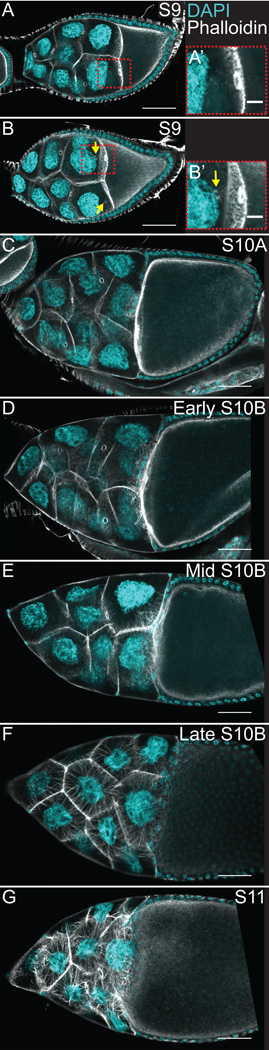
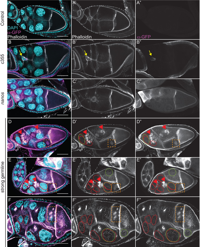
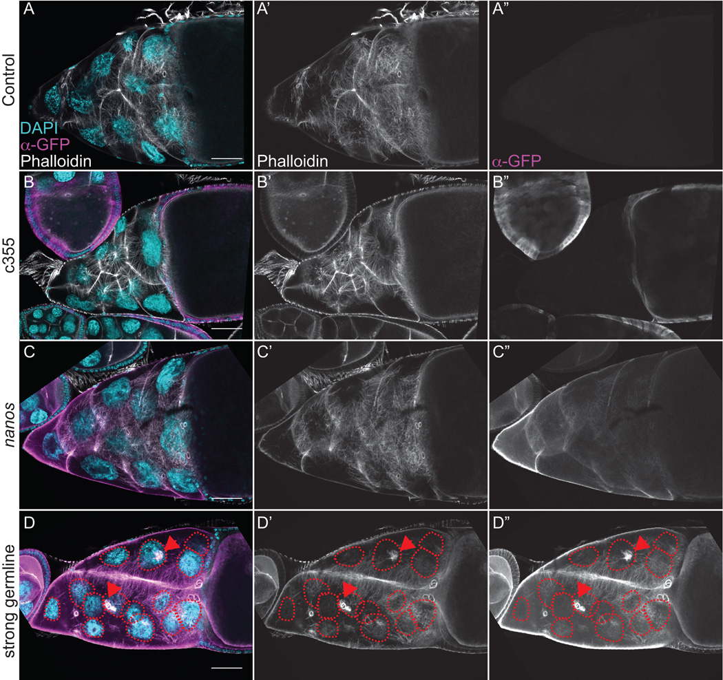
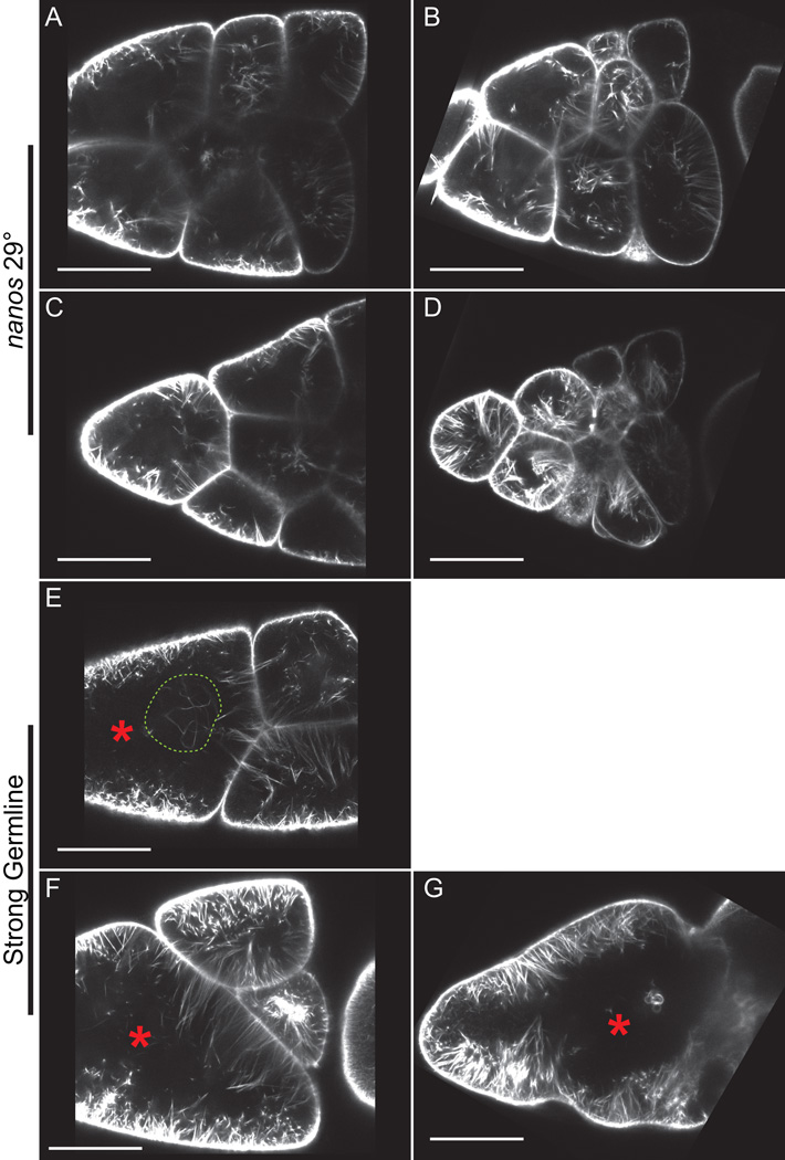
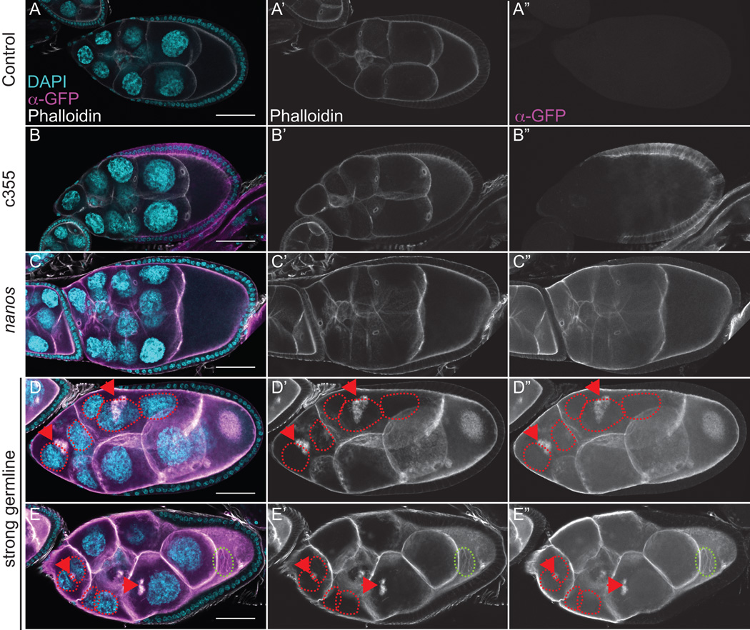
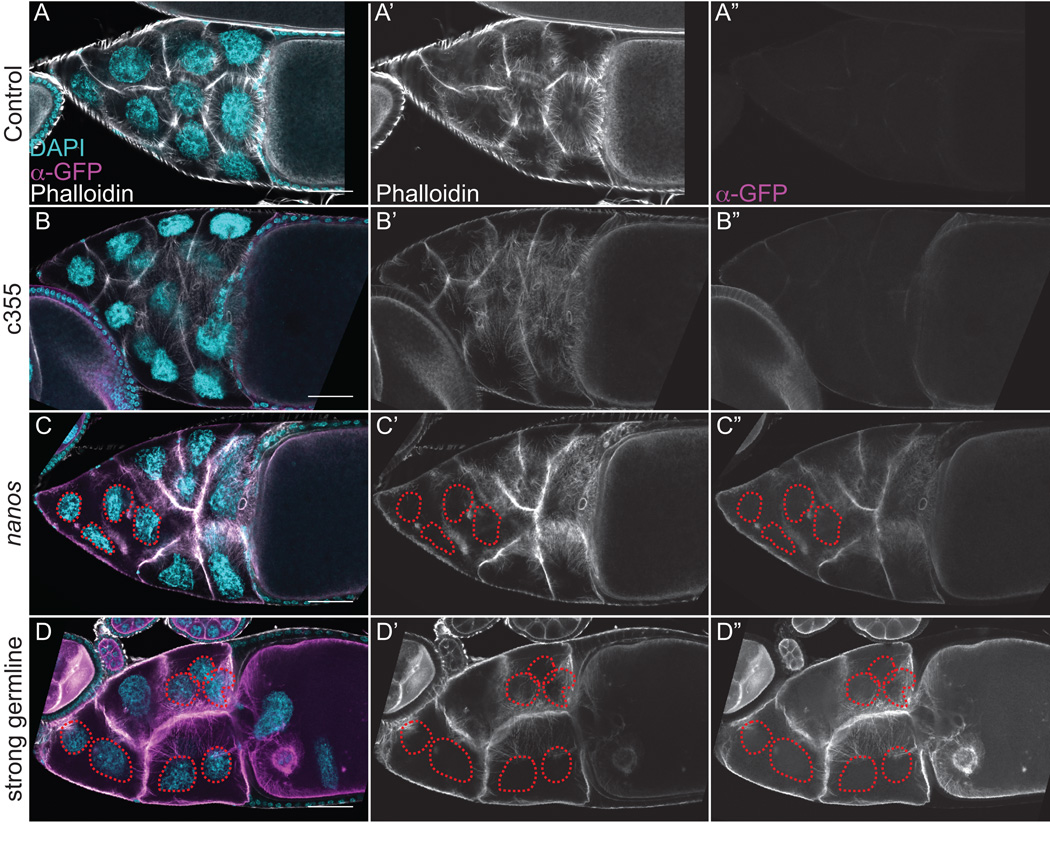
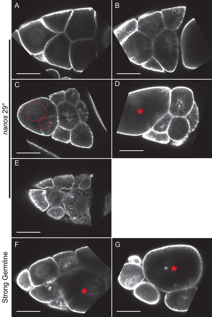
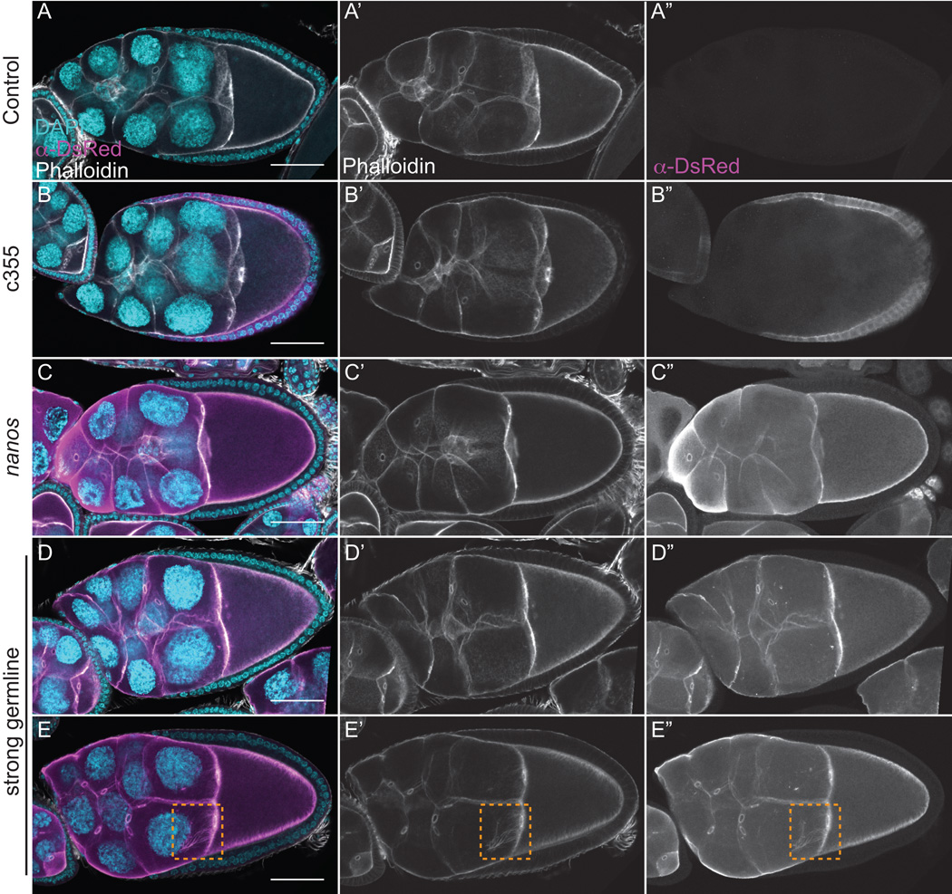
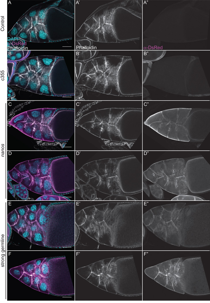
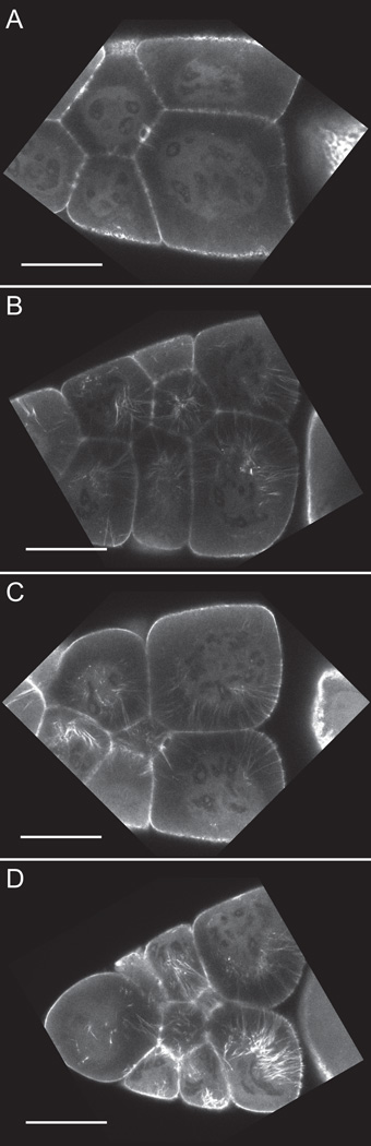
Dynamic remodeling of the actin cytoskeleton is required for both development and tissue homeostasis. While fixed image analysis has provided significant insight into such events, a complete understanding of cytoskeletal dynamics requires live imaging. Numerous tools for the live imaging of actin have been generated by fusing the actin-binding domain from an actin-interacting protein to a fluorescent protein. Here we comparatively assess the utility of three such tools--Utrophin, Lifeact, and F-tractin--for characterizing the actin remodeling events occurring within the germline-derived nurse cells during Drosophila mid-oogenesis or follicle development. Specifically, we used the UAS/GAL4 system to express these tools at different levels and in different cells, and analyzed these tools for effects on fertility, alterations in the actin cytoskeleton, and ability to label filamentous actin (F-actin) structures by both fixed and live imaging. While both Utrophin and Lifeact robustly label F-actin structures within the Drosophila germline, when strongly expressed they cause sterility and severe actin defects including cortical actin breakdown resulting in multi-nucleate nurse cells, early F-actin filament and aggregate formation during stage 9 (S9), and disorganized parallel actin filament bundles during stage 10B (S10B). However, by using a weaker germline GAL4 driver in combination with a higher temperature, Utrophin can label F-actin with minimal defects. Additionally, strong Utrophin expression within the germline causes F-actin formation in the nurse cell nuclei and germinal vesicle during mid-oogenesis. Similarly, Lifeact expression results in nuclear F-actin only within the germinal vesicle. F-tractin expresses at a lower level than the other two labeling tools, but labels cytoplasmic F-actin structures well without causing sterility or striking actin defects. Together these studies reveal how critical it is to evaluate the utility of each actin labeling tool within the tissue and cell type of interest in order to identify the tool that represents the best compromise between acceptable labeling and minimal disruption of the phenomenon being observed. In this case, we find that F-tractin, and perhaps Utrophin, when Utrophin expression levels are optimized to label efficiently without causing actin defects, can be used to study F-actin dynamics within the Drosophila nurse cells.

Keywords: Actin; Live Imaging; Oogenesis.


Figures

Figure 1
Figure 1. Actin remodeling is temporally and spatially regulated during mid-oogenesis
(A–G) Maximum projections of 3–5 confocal slices of fixed and stained wild-type (yw) follicles, staged as indicated, taken at 20X magnification. Anterior is to the left. F-actin (phalloidin) = white, DNA (DAPI) = cyan. (A-B’) S9. (C) S10A. (D-F) S10B. (G) S11. During S9, the border cells and main-body follicle cells are undergoing migration and the nurse cell cytoplasm is largely devoid of actin filament structures (A-A’), but occasionally there are actin filament and aggregate structures emanating from the ring canals in the posterior nurse cells adjacent to the oocyte (B-B’). Both the border cell and main-body follicle cell migration are completed by S10A and the nurse cells lack cytoplasmic F-actin structures (C). During S10B, dynamic actin remodeling is occurring within the nurse cells. Actin filament bundles first form in the posterior nurse cells, at the nurse cell-oocyte boundary (D). These bundles continue to elongate and bundle formation initiates on all of the nurse cell membranes that are directly attached to their neighboring nurse cell by a ring canal (E). At the completion of S10B the bundles are uniformly distributed along the nurse cell membranes and extend all the way to the nucleus (F). During S11, the cortical actin contracts to squeeze the cytoplasmic contents of the nurse cells into the growing oocyte (G). Scale bars = 50 μm, except in A’ and B’ where scale bars = 10 μm.
Figure 2
Figure 2. Strong germline expression of GFP-Utrophin results in cortical actin breakdown and the accumulation of F-actin in the nurse cell nuclei during S9
(A-E”) Maximum projections of 35 confocal slices of fixed and stained S9 follicles taken at 20X magnification. (F-F”) Maximum projections of 4–8 confocal slices of a fixed and stained S9 follicle taken at 63X magnification. (A-F) Merged images: DNA (DAPI) = cyan, F-actin (phalloidin) = white, Utrophin (anti-GFP) = magenta. (A’-F’) F-actin (phalloidin) = white. (A”-F”) Utrophin (anti-GFP) = white. (A-A”) GFP-Utrophin/+. (B-B”) c355 GAL4; GFP-Utrophin. (C-C”) GFP-Utrophin; nanos-VP16 GAL4. (D-F”) Strong germline GAL4 (either mat2MK, mat3, or oskar GAL4) driving GFP-Utrophin. Somatic expression of GFP-Utrophin has no effect on the nurse cell actin cytoskeleton or follicle cell morphology (B-B’’ compared to A-A’’), but expression within the border cells results in delayed migration (B-B”, yellow arrow). Weak germline expression of GFP-Utrophin is patchy and variable in the level of expression and ability to label cortical and ring canal F-actin, and does not alter F-actin morphology (C-C’’ compared to A-A’’). While cortical and ring canal F-actin label well when GFP-Utrophin is strongly expressed in the germline, high levels of GFP-Utrophin result in numerous defects including cortical actin breakdown with aggregates of ring canals (D-E”, red arrowheads and multi-nucleate nurse cells (D-F”, red circles), and early actin filaments and aggregates emanating from the ring canals in the posterior nurse cells (D-F”, orange boxes). Additionally, phalloidin and Utrophin positive F-actin structures that we term threads accumulate in the nurse cell nuclei and germinal vesicle when GFP-Utrophin is strongly expressed in the germline (E-F”, green circles). Scale bars = 50 μm.
Figure 3
Figure 3. GFP-Utrophin labels actin filament bundles during S10B, but strong germline expression results in cortical actin breakdown
(A-D”) Maximum projections of 3–5 confocal slices of fixed and stained S10B follicles taken at 20X magnification. (A-D) Merged images: DNA (DAPI) = cyan, F-actin (phalloidin) = white, Utrophin (anti-GFP) = magenta. (A’-D’) F-actin (phalloidin) = white. (A”-D”) Utrophin (anti-GFP) = white. (A-A”) GFP-Utrophin/+. (B-B”) c355 GAL4; GFP-Utrophin. (C-C”) GFP-Utrophin; nanos-VP16 GAL4. (D-D”) Strong germline GAL4 (either mat2MK, mat3, or oskar GAL4) driving GFP-Utrophin. Follicle cell morphology and nurse cell actin remodeling is not altered by either somatic expression or weak germline expression of GFP-Utrophin during S10B (B-B’’ and C-C”, respectively, compared to A-A’’). Both cytoplasmic actin filament bundles and cortical actin deposits are labeled by either weak or strong germline expression of GFP-Utrophin (C-D”). However, strong germline expression of GFP-Utrophin results in cortical actin breakdown with ring canal aggregates (red arrowheads) and multi-nucleate nurse cells (red circles), as well as elongated and disorganized actin bundles (D-D”). Scale bars = 50 μm.
Figure 4
Figure 4. Live imaging of S10B GFP-Utrophin expressing follicles
(A-G) Single focal plane from confocal z-stack of live S10B-S11 follicles taken at 40X magnification. (A-G) GFP-Utrophin = white. (A-D) GFP-Utrophin; nanos-VP16 GAL4 at 29°C. (E-G) Strong germline GAL4 (mat3 or oskar GAL4) driving GFP-Utrophin (room temperature). Cortical actin deposits and cytoplasmic actin filament bundles can be readily visualized when GFP-Utrophin is driven by nanos-VP16 GAL4 at 29° (A-D) or a strong germline GAL4 at room temperature (E-G). However, strong expression of GFP-Utrophin results in severe actin defects including cortical actin breakdown indicated by floating ring canals and oversized nurse cells (red asterisks, E-G), the formation of nuclear threads (E, dashed green circle), and disorganized bundles (F-G). Scale bars = 50 μm.
Figure 5
Figure 5. Strong germline expression of Lifeact-mEGFP results in cortical actin breakdown during S9
(A-E”) Maximum projections of 3–5 confocal slices of fixed and stained S9 follicles taken at 20X magnification. (A-E) Merged images: DNA (DAPI) = cyan, F-actin (phalloidin) = white, Lifeact (anti-GFP) = magenta. (A’-E’) F-actin (phalloidin) = white. (A”-E”) Lifeact (anti-GFP) = white. (A-A”) Lifeact-mEGFP /+. (B-B”) c355 GAL4; Lifeact-mEGFP. (C-C”) Lifeact-mEGFP; nanos-VP16 GAL4. (D-E”) Strong germline GAL4 (either mat2MK, mat3, or oskar GAL4) driving Lifeact-mEGFP. Neither somatic nor weak germline expression of Lifeact-mEGFP alters the nurse cell cytoskeleton or follicle cell morphology (B-B” and C-C”, respectively, compared to A-A”). Strong germline expression of Lifeact-mEGFP results in morphologic defects (D-E”), including misshapen nurse cells and oocyte, and cortical actin breakdown with aggregates of ring canals (red arrowheads) and multi-nucleate nurse cells (red circles). Additionally, phalloidin and Lifeact-mEGFP positive threads accumulate within the germinal vesicle following strong germline expression of Lifeact-mEGFP (E-E”, green circle). Scale bars = 50 μm.
Figure 6
Figure 6. Lifeact-mEGFP labels actin filament bundles during S10B, but germline expression results in cortical actin breakdown
(A-D”) Maximum projections of 3–5 confocal slices of fixed and stained S10B follicles taken at 20X magnification. (A-D) Merged images: DNA (DAPI) = cyan, F-actin (phalloidin) = white, Lifeact (anti-GFP) = magenta. (A’-D’) F-actin (phalloidin) = white. (A”-D”) Lifeact (anti-GFP) = white. (A-A”) Lifeact-mEGFP /+. (B-B”) c355 GAL4; Lifeact-mEGFP. (C-C”) Lifeact-mEGFP; nanos-VP16 GAL4. (D-D”) Strong germline GAL4 (either mat2MK, mat3, or oskar GAL4) driving Lifeact-mEGFP. Somatic expression of Lifeact-mEGFP does not alter nurse cell actin remodeling or follicle cell morphology during S10B (B-B” compared to A-A”). While Lifeact-mEGFP clearly labels cytoplasmic actin filament bundles within the nurse cells, both weak and strong germline expression of Lifeact-mEGFP results in cortical actin defects causing multi-nucleate nurse cells (C-D”, red circles). Scale bars = 50 μm.
Figure 7
Figure 7. Live imaging of S10B Lifeact-mEGFP expressing follicles
(A-G) Single focal plane from confocal z-stack of live S10B-S11 follicles taken at 40X magnification. (A-G) Lifeact-mEGFP = white. (A-D) Lifeact-mEGFP; nanos-VP16 GAL4 at 29°. (E-G) Strong germline GAL4 (mat3) driving Lifeact-mEGFP (room temperature). Cortical actin deposits and cytoplasmic actin filament bundles can be readily visualized when Lifeact-mEGFP is driven by nanos-VP16 GAL4 at 29° or a strong germline GAL4 at room temperature (A-G). Weakly driving expression of Lifeact-mEGFP fails to label actin filament bundle initiation (A), but does label actin bundle elongation (B), and contraction (C-D). Occasionally, cortical actin breakdown (C, nuclei within multi-nucleate nurse cell outlined with dashed red lines, and D, red asterisk) and disrupted actin filament bundle morphology (E) are observed under these conditions. In contrast, strong germline expression of Lifeact-mEGFP results in better labeling of F-actin structures, but also results in severe actin defects including cortical actin breakdown (red asterisks, F-G) and disorganized actin bundles (F-G). Scale bars = 50 μm.
Figure 8
Figure 8. F-tractin-tdTom labels F-actin structures during S9 without causing severe actin defects
(A-E”) Maximum projections of 3–5 confocal slices of fixed and stained S9 follicles taken at 20X magnification. (A-E) Merged images: DNA (DAPI) = cyan, F-actin (phalloidin) = white, F-tractin (anti-DsRed) = magenta. (A’-E’) F-actin (phalloidin) = white. (A”-E”) F-tractin (anti-DsRed)= white. (A-A”) F-tractin-tdTom/+. (B-B”) c355 GAL4; F-tractin-tdTom. (C-C”) F-tractin-tdTom; nanos-VP16 GAL4. (D-E”) Strong germline GAL4 (either mat2MK, mat3, or oskar GAL4) driving F-tractin-tdTom. Somatic expression of F-tractin-tdTom does not alter the nurse cell cytoskeleton or follicle cell morphology (B-B’’ compared to A-A’’). Similarly, follicles weakly expressing F-tractin-tdTom in the germline exhibit normal F-actin structures during S9 (C-C” compared to A-A”). While strong germline expression of F-tractin-tdTom does not cause morphologic defects (D-E”), it results in an increased frequency of early F-actin filaments in the posterior nurse cells (E-E”, orange box). Additionally, strong germline expression of F-tractin-tdTom results in the appearance of anti-DsRed positive, but phalloidin negative, puncta in the nurse cell cytoplasm (D”, E”). Scale bars = 50 μm.
Figure 9
Figure 9. F-tractin-tdTom labels F-actin structures during S10B without causing actin defects
(A-F”) Maximum projections of 3–5 confocal slices of fixed and stained S10B follicles taken at 20X magnification. (A-F) Merged images: DNA (DAPI) = cyan, F-actin (phalloidin) = white, F-tractin (anti-DsRed) = magenta. (A’-F’) F-actin (phalloidin) = white. (A”-F”) F-tractin (anti-DsRed)= white. (A-A”) F-tractin-tdTom/+. (B-B”) c355 GAL4; F-tractin-tdTom/+. (C-D”) F-tractin-tdTom/+; nanos-VP16 GAL4. (E-F”) Strong germline GAL4 (either mat2MK, mat3, or oskar GAL4) driving F-tractin-tdTom. Somatic expression of F-tractin-tdTom does not alter nurse cell actin remodeling or follicle cell morphology during S10B (B-B” compared to A-A”). Follicles weakly expressing F-tractin-tdTom in the germline exhibit normal nurse cell actin filament bundles and cortical actin deposits (C-D” compared to A-A”). However, the actin filament bundles within the nurse cells are unlabeled (C-C”) or only weakly labeled (D-D”) when F-tractin-tdTom is weakly expressed using nanos-VP16 GAL4. Strong germline expression of F-tractin-tdTom does not cause striking defects in nurse cell actin filament bundles and cortical actin deposits, and actin bundles are labeled, although to varying degrees (E-F”). Scale bars = 50 μm.
Figure 10
Figure 10. Live imaging of S10B F-tractin-tdTom expressing follicles
(A-D) Single focal plane from confocal z-stack of live S10B-S11 follicles taken at 40X magnification. (A-D) F-tractin-tdTom = white. (A-D) F-tractin-tdTom; mat3 GAL4 at 29°. Cortical actin deposits and cytoplasmic actin filament bundles can be readily visualized when F-tractin-tdTomato is driven using a strong germline GAL4 at 29°. Cytoplasmic actin filament bundle initiation (A), elongation (B-C), and contraction (D) are visualized. Scale bars = 50 μm.

Similar articles

Cited by

References

    1. Abramoff MD, Magalhaes PJ, Ram SJ. Image Processing with Image. J. Biophotonics International. 2004;11:36–42.
    1. Aizawa H, Sameshima M, Yahara I. A green fluorescent protein-actin fusion protein dominantly inhibits cytokinesis, cell spreading, and locomotion in Dictyostelium. Cell structure and function. 1997;22:335–345. - PubMed
    1. Baarlink C, Wang H, Grosse R. Nuclear actin network assembly by formins regulates the SRF coactivator MAL. Science. 2013;340:864–867. - PubMed
    1. Belin BJ, Cimini BA, Blackburn EH, Mullins RD. Visualization of actin filaments and monomers in somatic cell nuclei. Molecular biology of the cell. 2013;24:982–994. - PMC - PubMed
    1. Boyle MJ, French RL, Cosand KA, Dorman JB, Kiehart DP, Berg CA. Division of labor: subsets of dorsal-appendage-forming cells control the shape of the entire tube. Developmental biology. 2010;346:68–79. - PMC - PubMed

Publication types

LinkOut - more resources