. 2014 Jul 3;95(1):121-5.
doi: 10.1016/j.ajhg.2014.06.007.

Aicardi-Goutières syndrome is caused by IFIH1 mutations

Affiliations

Aicardi-Goutières syndrome is caused by IFIH1 mutations

Hirotsugu Oda et al. Am J Hum Genet. .

Abstract

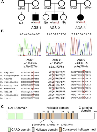
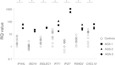
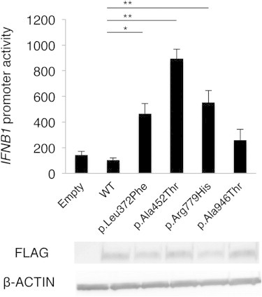
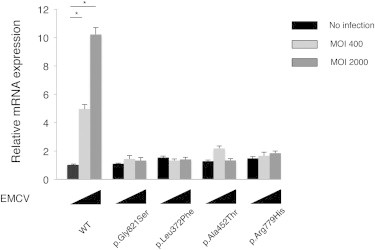
Aicardi-Goutières syndrome (AGS) is a rare, genetically determined early-onset progressive encephalopathy. To date, mutations in six genes have been identified as etiologic for AGS. Our Japanese nationwide AGS survey identified six AGS-affected individuals without a molecular diagnosis; we performed whole-exome sequencing on three of these individuals. After removal of the common polymorphisms found in SNP databases, we were able to identify IFIH1 heterozygous missense mutations in all three. In vitro functional analysis revealed that IFIH1 mutations increased type I interferon production, and the transcription of interferon-stimulated genes were elevated. IFIH1 encodes MDA5, and mutant MDA5 lacked ligand-specific responsiveness, similarly to the dominant Ifih1 mutation responsible for the SLE mouse model that results in type I interferon overproduction. This study suggests that the IFIH1 mutations are responsible for the AGS phenotype due to an excessive production of type I interferon.


Figures

Figure 1
Figure 1
Pedigree Information for the AGS-Affected Individuals and Details of the IFIH1 Mutations Identified (A) The pedigrees of the three families indicating the AGS probands. (B) Sanger sequencing chromatograms of the three IFIH1 mutations found in the AGS-affected individuals. The locations of these mutations in the amino acid sequence of the MDA5 protein are shown in alignment with the conserved amino acid sequences from several species. This alignment was obtained via ClustalW2. The amino acids that are conserved with human are circled in red. (C) The MDA5 protein domain structure with the amino acid substitutions observed in these AGS-affected individuals.
Figure 2
Figure 2
Quantitative RT-PCR of a Panel of Seven ISGs in PBMCs Obtained from the IFIH1-Mutated Individuals and Healthy Control Subject qRT-PCR was performed as previously described. The relative abundance of each transcript was normalized to the expression level of β-actin. Taqman probes used were the same as previous report, except for ACTB (MIM 102630). Individual data were shown relative to a single calibrator (control 1). The experiment was performed in triplicate. Statistical significance was determined by Mann-Whitney U test, p < 0.05.
Figure 3
Figure 3
The Effects of the Three MDA5 Variants on IFNB1 Expression Huh7 cells were transfected with a reporter gene containing IFNB1 promoter (p-55C1B Luc), an empty vector (BOS), and expression vectors for FLAG-tagged human wild-type IFIH1, c.2836G>A polymorphism (p.Ala946Thr) in the GWASs, and the identified IFIH1 mutants. Luciferase activity was measured 48 hr after transfection, and the MDA5 protein accumulation was examined by immunoblotting as previously described. FLAG indicates the accumulation of FLAG-tagged MDA5. Each experiment was performed in triplicate and data are mean ± SEM. Shown is a representative of two with consistent results. Statistical significance was determined by Student’s t test. p < 0.05, ∗∗p < 0.01.
Figure 4
Figure 4
Ifnb mRNA Levels in Ifih1-Deficient MEFs Expressing IFIH1 Mutants The MEFs were infected with retroviruses encoding mouse wild-type Ifih1, mouse Ifih1 with c.2461G>A (p.Gly821Ser) (RefSeq NM_027835.3) mutation, or the three AGS mutants of human IFIH1. At 48 hr after the retroviral infection, these MEFs were infected with indicated multiplicity of infection (MOI) of EMCV for 6 hr, and Ifnb mRNA levels were measured by qRT-PCR. The relative abundance of each transcript was normalized to the expression level of 18S ribosomal RNA. Data are shown as mean ± SEM of triplicate samples. Shown is a representative of two independent experiments. Statistical significance was determined by Student’s t test, p < 0.001. The expression of the retrovirally transduced FLAG-tagged constructs was confirmed by immunoblotting (Figure S5).

Comment in

Similar articles

Cited by

References

    1. Chahwan C., Chahwan R. Aicardi-Goutieres syndrome: from patients to genes and beyond. Clin. Genet. 2012;81:413–420. - PubMed
    1. Ramantani G., Kohlhase J., Hertzberg C., Innes A.M., Engel K., Hunger S., Borozdin W., Mah J.K., Ungerath K., Walkenhorst H. Expanding the phenotypic spectrum of lupus erythematosus in Aicardi-Goutières syndrome. Arthritis Rheum. 2010;62:1469–1477. - PubMed
    1. Orcesi S., La Piana R., Fazzi E. Aicardi-Goutieres syndrome. Br. Med. Bull. 2009;89:183–201. - PubMed
    1. Rice G., Patrick T., Parmar R., Taylor C.F., Aeby A., Aicardi J., Artuch R., Montalto S.A., Bacino C.A., Barroso B. Clinical and molecular phenotype of Aicardi-Goutieres syndrome. Am. J. Hum. Genet. 2007;81:713–725. - PMC - PubMed
    1. Blau N., Bonafé L., Krägeloh-Mann I., Thöny B., Kierat L., Häusler M., Ramaekers V. Cerebrospinal fluid pterins and folates in Aicardi-Goutières syndrome: a new phenotype. Neurology. 2003;61:642–647. - PubMed

Publication types

Substances

Supplementary concepts