Review
. 2014 Jul 3;15(1):14-25.
doi: 10.1016/j.stem.2014.06.009.

Immune modulation of stem cells and regeneration

Affiliations
Review

Immune modulation of stem cells and regeneration

Arin B Aurora et al. Cell Stem Cell. .

Abstract

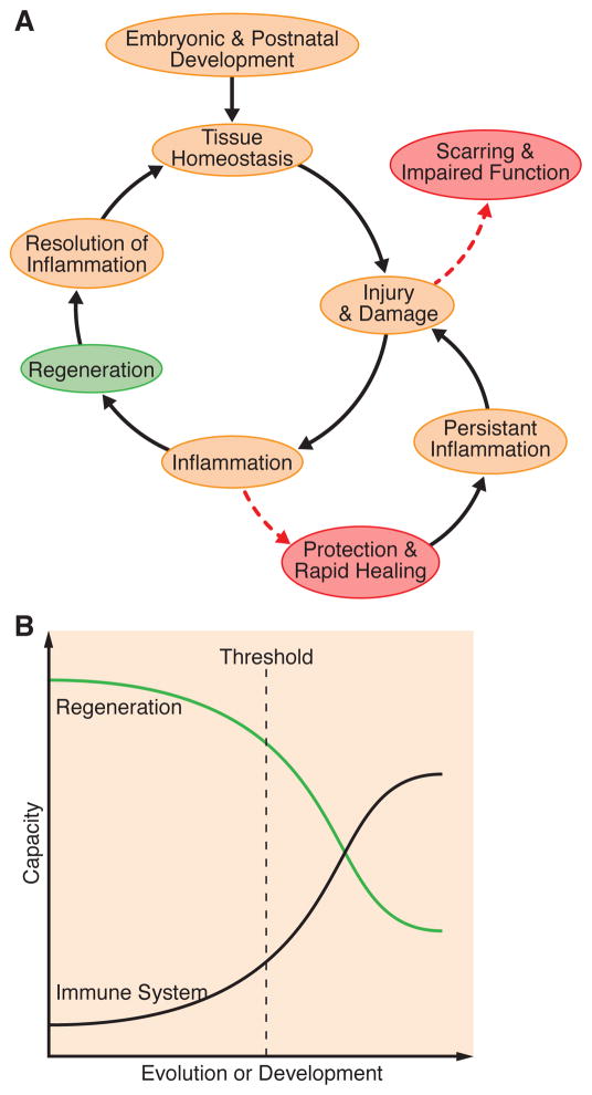
The immune system, best known as the first line of defense against invading pathogens, is integral to tissue development, homeostasis, and wound repair. In recent years, there has been a growing appreciation that cellular and humoral components of the immune system also contribute to regeneration of damaged tissues, including limbs, skeletal muscle, heart, and the nervous system. Here, we discuss key findings that implicate inflammatory cells and their secreted factors in tissue replacement after injury via stem cells and other reparative mechanisms. We highlight clinical conditions that are amenable to immune-mediated regeneration and suggest immune targeting strategies for tissue regeneration.


Figures

Figure 1
Figure 1. The influence of the immune system on development, homeostasis and disease
(A) During embryonic and postnatal development, the immune system regulates processes such as branching morphogenesis, ductal formation and angiogenesis. Similar functions are maintained in some adult tissues to maintain normal homeostasis. Injury or disease elicits an inflammatory response that can either promote functional restoration of the tissue (regeneration) or a rapid healing response that may protect the organism at the expense of preserving structure and function. Inflammation usually resolves in the regenerative response while inflammation often persists in wound healing and scar formation, ultimately impairing the normal function of the tissue. (B) Inverse relationship between the capacity to regenerate and the strength and intricacy of the immune system during development or evolution. The threshold indicated on the graph conceptualizes the balance point at which the pro-regenerative components of the immune response are maintained within the context of a more advanced immune system. Identifying this threshold will be a key step towards the development of regenerative therapies targeted at immunity.
Figure 2
Figure 2. Debris clearance as a coordinator of regeneration
Debris clearance orchestrated by the immune system is a key activator of subsequent steps in regeneration, including progenitor cell activation, differentiation, and immune polarization. The comparison shown between skeletal muscle and the CNS highlights the key cell types and soluble factors involved. Following skeletal muscle injury, both M1 macrophages and fibro/adipocyte progenitors (FAPs) clear cellular debris. FAP phagocytic activity depends on eosinophil derived IL-4; in its absence, the progenitors differentiate into fat, causing muscle dysfunction. Phagocytic M1 macrophages promote myoblast proliferation and polarize to an M2 phenotype via AMPK-mediated signaling. M2 polarization is required for appropriate myoblast differentiation. In the CNS, mature neurons lack robust regenerative potential. However, remyelination occurs in young, healthy adults by activation of oligodendrocyte progenitor cells (OPCs). Activation of OPC proliferation depends on efficient clearance of myelin debris, which contains inhibitory factors, and also macrophage -derived soluble factors. Similar to myoblasts, the proliferation and recruitment of OPCs depends on M1 macrophages while differentiation of OPCs and remyelination relies on M2 macrophage-secreted activin-A.
Figure 3
Figure 3. Immune cell polarization and heterogeneity are key components of tissue repair or regeneration
(A) The regenerative capacity of skeletal muscle is driven by satellite cell activation, proliferation and differentiation. M1 macrophages are early responders that secrete cytokines with proliferative effects on myoblasts. The next phase of the response involves myotube differentiation driven by M2 macrophage-secreted IGF1 and TGFβ. Specialized muscle Treg cells influence all stages of regeneration: early secretion of amphiregulin activates myoblast proliferation, while in subsequent phases of regeneration muscle Treg are necessary for myotube differentiation, M1 to M2 polarization and attenuation of excessive T lymphocyte responses. (B) The adult mammalian heart, which lacks regenerative capacity, contains different cardiac macrophage subsets, with diverse functions, developmental origins, and mechanisms of homeostasis. The four populations, segregated by expression levels of CCR2, Ly-6C, and MHC class II, perform different functions as indicated. Upon injury such as a MI, a biphasic monocyte response occurs to promote an initial inflammatory phase followed by a reparative phase mediated by Ly-6Chi or Ly-6Clo splenic monocytes respectively. The two systems are linked by the ability of splenic Ly-6Chi monocytes to replenish all four subsets in Phase I. In the neonatal mouse heart, which can fully regenerate following MI, there is also a biphasic splenic monocyte response though the characterization of subsets of resident cardiac macrophages has yet to be investigated. Interestingly, neonatal cardiac macrophages differ from adult in their localization, abundance, and gene expression profile following injury and are required for regeneration by promoting angiogenesis. The regenerative subtype in the neonate has yet to be defined.

References

    1. Arnold L, Henry A, Poron F, Baba-Amer Y, van Rooijen N, Plonquet A, Gherardi RK, Chazaud B. Inflammatory monocytes recruited after skeletal muscle injury switch into antiinflammatory macrophages to support myogenesis. J Exp Med. 2007;204:1057–1069. - PMC - PubMed
    1. Aurora AB, Porrello ER, Tan W, Mahmoud AI, Hill JA, Bassel-Duby R, Sadek HA, Olson EN. Macrophages are required for neonatal heart regeneration. J Clin Invest. 2014;124:1382–1392. - PMC - PubMed
    1. Baeyens L, Lemper M, Leuckx G, De Groef S, Bonfanti P, Stange G, Shemer R, Nord C, Scheel DW, Pan FC, et al. Transient cytokine treatment induces acinar cell reprogramming and regenerates functional beta cell mass in diabetic mice. Nature biotechnology. 2014;32:76–83. - PMC - PubMed
    1. Banaei-Bouchareb L, Gouon-Evans V, Samara-Boustani D, Castellotti MC, Czernichow P, Pollard JW, Polak M. Insulin cell mass is altered in Csf1op/Csf1op macrophage-deficient mice. J Leukoc Biol. 2004;76:359–367. - PubMed
    1. Barrette B, Hebert MA, Filali M, Lafortune K, Vallieres N, Gowing G, Julien JP, Lacroix S. Requirement of myeloid cells for axon regeneration. J Neurosci. 2008;28:9363–9376. - PMC - PubMed

Publication types