Review
. 2014 Sep:74:157-74.
doi: 10.1016/j.freeradbiomed.2014.06.006. Epub 2014 Jul 1.

The 2013 SFRBM discovery award: selected discoveries from the butterfield laboratory of oxidative stress and its sequela in brain in cognitive disorders exemplified by Alzheimer disease and chemotherapy induced cognitive impairment

Affiliations
Review

The 2013 SFRBM discovery award: selected discoveries from the butterfield laboratory of oxidative stress and its sequela in brain in cognitive disorders exemplified by Alzheimer disease and chemotherapy induced cognitive impairment

D Allan Butterfield. Free Radic Biol Med. 2014 Sep.

Abstract

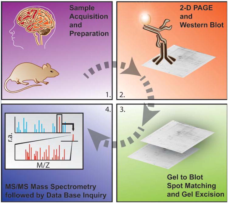
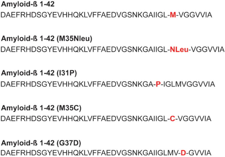
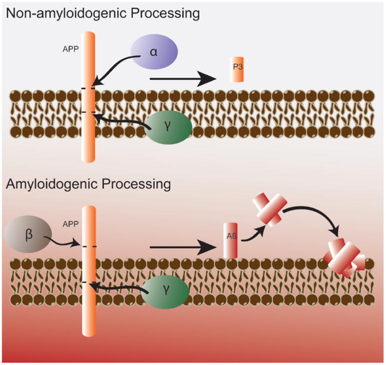
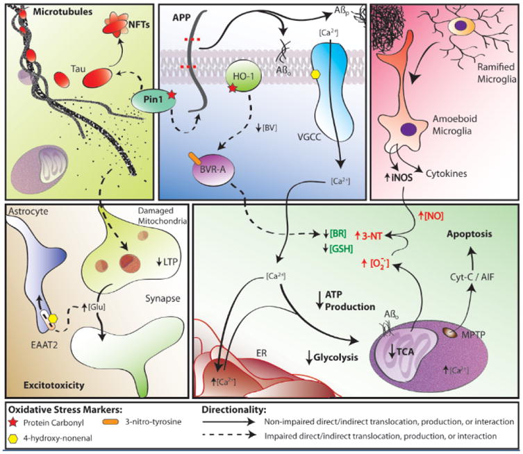
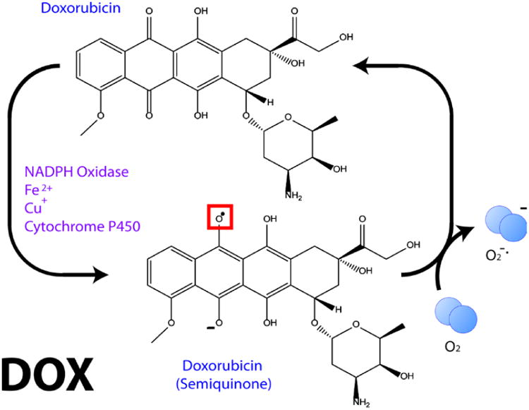
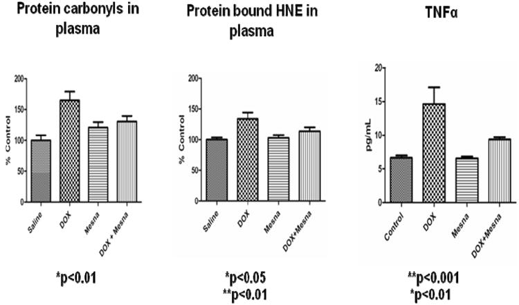
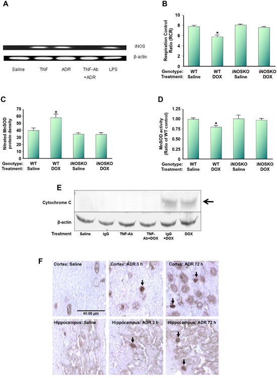
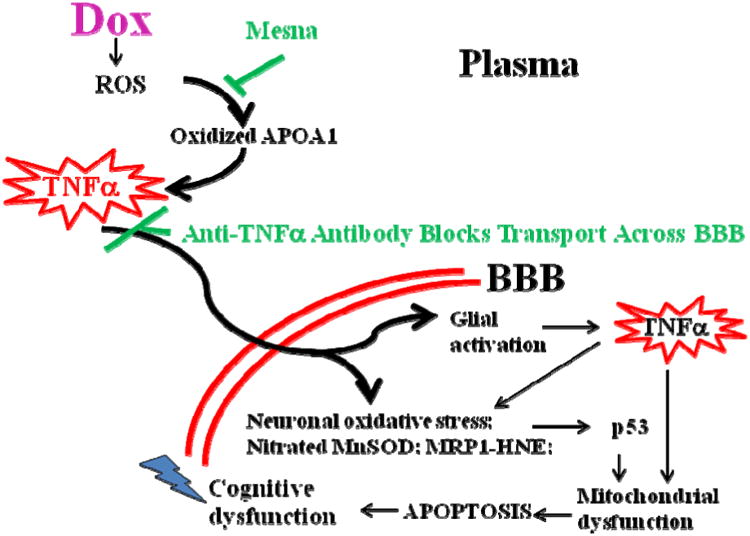
This retrospective review on discoveries of the roles of oxidative stress in brain of subjects with Alzheimer disease (AD) and animal models thereof as well as brain from animal models of chemotherapy-induced cognitive impairment (CICI) results from the author receiving the 2013 Discovery Award from the Society for Free Radical Biology and Medicine. The paper reviews our laboratory's discovery of protein oxidation and lipid peroxidation in AD brain regions rich in amyloid β-peptide (Aβ) but not in Aβ-poor cerebellum; redox proteomics as a means to identify oxidatively modified brain proteins in AD and its earlier forms that are consistent with the pathology, biochemistry, and clinical presentation of these disorders; how Aβ in in vivo, ex vivo, and in vitro studies can lead to oxidative modification of key proteins that also are oxidatively modified in AD brain; the role of the single methionine residue of Aβ(1-42) in these processes; and some of the potential mechanisms in the pathogenesis and progression of AD. CICI affects a significant fraction of the 14 million American cancer survivors, and due to diminished cognitive function, reduced quality of life of the persons with CICI (called "chemobrain" by patients) often results. A proposed mechanism for CICI employed the prototypical ROS-generating and non-blood brain barrier (BBB)-penetrating chemotherapeutic agent doxorubicin (Dox, also called adriamycin, ADR). Because of the quinone moiety within the structure of Dox, this agent undergoes redox cycling to produce superoxide free radical peripherally. This, in turn, leads to oxidative modification of the key plasma protein, apolipoprotein A1 (ApoA1). Oxidized ApoA1 leads to elevated peripheral TNFα, a proinflammatory cytokine that crosses the BBB to induce oxidative stress in brain parenchyma that affects negatively brain mitochondria. This subsequently leads to apoptotic cell death resulting in CICI. This review outlines aspects of CICI consistent with the clinical presentation, biochemistry, and pathology of this disorder. To the author's knowledge this is the only plausible and self-consistent mechanism to explain CICI. These two different disorders of the CNS affect millions of persons worldwide. Both AD and CICI share free radical-mediated oxidative stress in brain, but the source of oxidative stress is not the same. Continued research is necessary to better understand both AD and CICI. The discoveries about these disorders from the Butterfield Laboratory that led to the 2013 Discovery Award from the Society of Free Radical and Medicine provide a significant foundation from which this future research can be launched.

Keywords: 2013 SFRBM Discovery Award; Alzheimer disease (AD) and its earlier forms (amnestic MCI and preclinical AD); Aβ(1–42) associated oxidative stress; Chemotherapy-induced cognitive impairment (“chemobrain”); Plasma-derived elevated TNFα and its sequela in brain; Redox proteomics.


Figures

Figure 1
Figure 1
Schematic representation of the major methodological steps involved in redox proteomics. For more details see recent reviews in [6, 7]. The figure, used with permission from Elsevier Science Publishers, is taken from reference [122].
Figure 2
Figure 2
Sequences of Aβ(1-42) and variants of this sequence mentioned in this retrospective paper. Note the key methionine residue at position 35 of Aβ(1-42).
Figure 3
Figure 3
Non-amyloidogenic (Top) and amyloidogenic (Bottom) pathways of APP processing. Note the latter leads to olgimeric Aβ(1-42) that enters the lipid bilayer leading to damaging lipid peroxidation and its sequelae as discussed in the text. The figure, used with permission from Elsevier Science Publishers, is taken from reference [122].
Figure 4
Figure 4
A,B. Structures of antioxidant compounds mentioned in this retrospective paper.
Figure 4
Figure 4
A,B. Structures of antioxidant compounds mentioned in this retrospective paper.
Figure 5
Figure 5
N-Acetylcysteine (NAC) given in drinking water inhibits oxidative stress in brain at 9 mos. of age of the APP/PS-1 human double mutant knock in mouse model of AD. Protein oxidation in brain assessed by protein carbonyls is shown. The figure is adapted from [91].
Figure 6
Figure 6
A,B. Role of the single methionine residue of Aβ(1-42) in lipid peroxidation, whose mechanism is shown. Note the HNE formed by lipid peroxidation forms Michael adducts with protein-resident Cys, His, or Lys residues, changing the conformation and decreasing function of the HNE-modified proteins.
Figure 6
Figure 6
A,B. Role of the single methionine residue of Aβ(1-42) in lipid peroxidation, whose mechanism is shown. Note the HNE formed by lipid peroxidation forms Michael adducts with protein-resident Cys, His, or Lys residues, changing the conformation and decreasing function of the HNE-modified proteins.
Figure 7
Figure 7
Some findings and discoveries related to Aβ(1-42) oligomer-induced oxidative stress and its sequela published from the Butterfield laboratory that contribute to a unifying oxidative stress centric hypothesis for neuronal death in AD that is consistent with the pathology, biochemical alterations, and clinical presentation in this disorder. Amyloid β-peptide, produced by β- and γ-secretase (top, middle panel), aggregates into extracellular fibrils forming the core of SP or aggregates into hydrophobic oligomers that insert into the plasma membrane. One-electron oxidation of the S-atom of Met-35 of Aβ(1-42) [see text] initates the chain reaction of lipid peroxidation (LPO), greatly amplyfing the damage of the initial free radical on the peptide. HNE, produced by LPO, binds to and causes dysfunction of key proteins in the PM and cytosol. Among the former are ion-motive ATPases, e.g., Na,K-ATPase and Ca-ATPase. The resultant loss of cell potential opens voltage gated Ca2+ channels, leading to a massive influx of Ca2+ to the cytosol (bottom, right panel), and subsequent attempts to sequester this Ca2+ in ER and mitochondria. However, this massive overload of Ca2+ causes ER to undergo unfolded protein response and damage and decreases protein synthesis. Mitochondrial Ca2+ overload causes swelling of mitochondria and opening of the MPTP with release of cytochrome c to initiate the intrinsic pathway for apoptosis (bottom, right panel). The intracellular Ca2+ also activates numerous degradative enzymes such as calpains, PLA2, endonucleases, etc. causing necrotic mechanisms to engage (see text). The oxidative stress in cytosol and mitochondria lead to damage to many glycolytic, TCA, and ETC enzymes or complexes, resulting in dramatic loss of ATP production (bottom, right panel), which leads to loss of many important neuronal functions, ranging from axonal transport to neurotransmission (bottom, left panel), as well as maintaining the cellular and organelle potentials. This oxidative stress also damages Pin1, which regulates both APP and Tau (top, left/middle panels), but also a key tau kinase (GSK-3β) and a tau phosphatase (PP2A). The resultant hyperphosphorylated tau, falls off microtubules (MT) [top, left panel], leading to cessation of anterograde and retrograde transport. Among other determimental consequences of this, synaptic-resident mitochondria are no longer able to produce the required ATP to maintain presynaptic function, including loss of LTP, needed for learning and memory (bottom, left panel). Note that Pin1 is involved in three major neuropathological hallmarks of AD: SP, NFT, and synapse loss. The intracellular detritus resulting from all these aberrant processes is not removed, since the proteostasis network of the ubiquitin-proteasome system (UPS), ER, and autophagy are all damaged by oxidative stress (see text). The Aβ(1-42)-initiated oxidative stress also leads to damage to heme oxygenase -1 (HO-1) and biliverdin reductase-A (BVR-A), which decreases intracellular antioxidant bilirubin (top, middle panel; bottom, right panel). Moreover, glutathione (GSH) is decreased because of elevated oxidative stress and since its rate limiting synthetic enzyme, γ-Glu-Cys ligase, is damaged by oxidative stress. There are many other processes associated with oxidative stress on which we and others have published. However, this schematic diagram outlines some of the key processes involved in neuronal death in AD that fit the pathology, biochemical alterations, and clinical presentations observed in AD that were published from our laboratory based on neurochemical, oxidative stress, and proteomics studies of AD and its earlier forms.
Figure 8
Figure 8
Redox cycling of Dox to produce superoxide free radical. See text for more details.
Figure 9
Figure 9
Dox administration to WT mice (i.p.) leads in plasma to protein oxidation and lipid peroxidation (assessed by protein carbonyls and protein-bound HNE, respectively), and to elevated TNFα. Concomitant administration of Mesna significantly depresses these markers to essentially control levels. The figure, used with permission from Elsevier Science Publishers, is taken from reference [174].
Figure 10
Figure 10
Dox (also called adriamycin, ADR) induces nitrosative stress in brain mitochondria. WT mice were injected i.p. with Dox. (A). i-NOS message was induced, but anti-TNFα antibody prevents i-NOS induction following ADR treatment. (B). The RCR, a measure of oxygen consumption, in brain mitochondria isolated from ADR-treated WT mice is significantly depressed, but not in i-NOS knock-out mice. (C). Mitochondrial-resident MnSOD is nitrated following ADR addition to WT mice, but not in i-NOS knock-out mice. (D). MnSOD activity is significantly depressed in mitochondria isolated from brain of ADR-treated WT mice, but not in i-NOS knock-out mice. (E). Mitochondria isolated from brain of ADR-treated WT mice lead to cytochrome c release, but not in mice also treated with anti-TNFα antibody. Note that ADR-treated WT mice also treated with IgG still lead to cytochrome c release from mitochondria isolated from brain, suggesting specificity of the anti-TNFα treatment. (F). Consistent with the results of (E), apoptosis occurs in brain of ADR-treated WT mice as assessed by TUNEL staining even at 3h post-ADR treatment, and pronounced apoptosis at 72h post-ADR treatment occurred. This latter time is the time at which oxidative stress in brain is maximal. The figure, used with permission from Elsevier Science Publishers, was modified from reference [171].
Figure 11
Figure 11
Model for CICI (“chemobrain”) based on demonstrated results from our laboratory. Dox (ADR), as a prototypical ROS-generating agent that does not cross the BBB, causes oxidation of ApoA1 in plasma, which leads to increased levels of TNFα. This cytokine crosses the BBB to enter glia and neurons, generating even more TNFα. Subsequent biochemical events lead to brain mitochondrial dysfunction and resultant apoptosis as explained more in the text. Mesna, which does not interfere with cancer chemotherapy, blocks Dox-mediated oxidation of ApoA1 and elevation of TNFα. Anti- TNFα antibody blocks elevated TNFα from crossing the BBB. Both treatments inhibit the biochemical events that otherwise cause neuronal death. See text and [169-171, 174, 176, 181, 182] for more details.

References

    1. Halliwell B, Gutteridge J. Free Radicals in Biology and Medicine. Oxford University Press; 2007. p. 704.
    1. Butterfield DA, Stadman ER. Protein oxidation processes in aging brain. Advances in Cell Aging and Gerontology. 1997;2:161–191.
    1. Beckman JS, Chen J, Ischiropoulos H, Crow JP. Oxidative chemistry of peroxynitrite. Methods in enzymology. 1994;233:229–240. - PubMed
    1. Butterfield DA, Bader Lange ML, Sultana R. Involvements of the lipid peroxidation product, HNE, in the pathogenesis and progression of Alzheimer's disease. Biochim Biophys Acta. 2010;1801:924–929. - PMC - PubMed
    1. Esterbauer H, Schaur RJ, Zollner H. Chemistry and biochemistry of 4-hydroxynonenal, malonaldehyde and related aldehydes. Free Radic Biol Med. 1991;11:81–128. - PubMed

MeSH terms