. 2014 Aug;5(4):483-92.
doi: 10.1007/s13244-014-0338-0. Epub 2014 Jul 5.

Pulmonary Langerhans cell histiocytosis: the many faces of presentation at initial CT scan

Affiliations

Pulmonary Langerhans cell histiocytosis: the many faces of presentation at initial CT scan

M C Castoldi et al. Insights Imaging. 2014 Aug.

Abstract

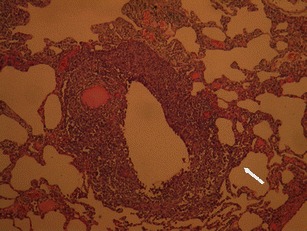
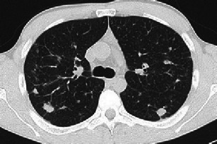
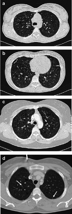
Objectives: Pulmonary Langerhans cell histiocytosis (PLCH) is a rare interstitial granulomatous disease that usually affects young adults who are smokers. Chest computed tomography (CT) allows a confident diagnosis of PLCH only in typical presentation, when nodules, cavitated nodules and cysts coexist and predominate in the upper and middle lungs.

Methods: This article includes a pictorial essay of typical and atypical presentations of PLCH at initial chest CT. Various appearances of PLCH are illustrated and possible differential diagnosis is discussed.

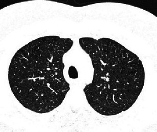
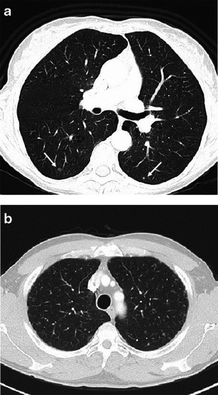
Results: PLCH can present with some aspecific features that may cause diagnosis of the initial disease to be overlooked or other pulmonary diseases to be suspected. In cases of nodule presentation alone, the main differential diagnosis should include lung metastasis, tuberculosis and other infections, sarcoidosis, silicosis and Wegener's disease. In cases of cysts alone, the most common diseases to be differentiated are centrilobular emphysema and lymphangiomyomatosis. Clinical symptoms are usually non-specific, although a history of cigarette smoking, coupled with the presence of typical or suggestive findings at imaging, is key to suspecting the disease. Atypical presentations require surgical biopsy for diagnosis.

Conclusions: The radiologist should be familiar with PLCH imaging features to correctly diagnose the disease or need for further investigation.

Teaching points: • PLCH is a rare interstitial smoking-related disease that usually affects young adults. • The typical first CT shows a mix of nodules, cavitary nodules and cysts in the upper-middle lungs. • Atypical appearance, either cysts or nodules alone, mandates that other diagnoses be considered. • Lung cystic involvement correlates with lung function abnormalities and predicts functional decline. • Integration of the clinical history and imaging results is key to diagnosis.


Figures

Fig. 1
Fig. 1
A 20-year-old woman, non-smoker, with PLCH; pulmonary cysts at her first computed tomography (CT) examination. The patient was treated in the neonatal period for a cutaneous form of Langerhans cell histiocytosis that regressed completely during infancy. The cysts are postulated to have resulted from granolomatous transformation of pulmonary histiocytosis, possibly concurrent with the cutaneous paediatric disease. Cysts remained stable during 10 years of follow-up
Fig. 2
Fig. 2
Low-power photomicrograph (original magnification, haematoxylin and eosin stain) of lung tissue shows dense non-necrotising granulomatous infiltration. The sample demonstrates well both localisation of the lesion around a dilated terminal airway and bronchial wall destruction by the infiltrate. The inflammatory cell infiltration also shows an interstitial distribution (arrow) (courtesy of Dr. E. Bonacina)
Fig. 3
Fig. 3
A 41-year-old woman with PLCH. Coronal CT image (thin MIP reconstruction) showing typical distribution of disease: many cysts predominating in upper and middle lungs fiends, with some micronodules (arrow); typically, basal regions are spared
Fig. 4
Fig. 4
Different imaging presentations of PLCH at clinical onset. Definitive diagnosis obtained by VATS performed within a month of imaging. a A 48-year-old woman, smoker, symptomatic for dry cough and mild fever. A 3-mm collimated CT shows a pattern of early “florid” disease characterised by numerous scattered nodules, of which almost all are cavitated and surrounded by ground glass opacities. A consolidation area also coexists (arrow). b A 46-year-old woman, symptomatic for dry cough and mild fever. HRCT scans show diffuse ill-defined micronodularity, cavitating nodules, cysts and mild interstitial thickening. c A 50-year-old woman, smoker, symptomatic for dry cough. HRCT scans show cystic disease with a few scattered micronodules
Fig. 5
Fig. 5
A 34-year-old man, smoker, symptomatic for cough and fever. Definitive diagnosis obtained by VATS performed within a month of imaging. A 3-mm collimated CT shows scattered nodules, with asymmetric distribution. The largest nodules measure 15–20 mm; nodular borders are irregular or atypically smooth. Smooth linear interstitial thickening is also evident
Fig. 6
Fig. 6
A 38-year-old man, non-smoker, with asymptomatic PLCH and skin involvement. Diagnosis obtained by skin lesion biopsy. BAL >5 % CD1a + cells. HRCT scans at diagnosis show a tiny micronodularity along with multiple areas of centrilobular emphysema
Fig. 7
Fig. 7
PLCH presenting with nodular features that mimic other diseases. a PLCH referred to metastasis: a 44-year-old woman, smoker, with a medical history of carcinoma of cervix uteri, treated with surgery and chemotherapy. A 3-mm collimated CT performed during oncological follow-up shows some faint micronodules in both lungs (arrows). Definitive diagnosis of PLCH obtained by VATS. b PLCH mimicking sarcoidosis: a 41-year-old man, smoker, with asymptomatic PLCH and skull involvement. Diagnosis obtained by skull biopsy. A 3-mm collimated CT shows numerous millimetric nodules. Nodules are mostly spread at the upper and middle lung fields in a peripheral location; few are subpleuric
Fig. 8
Fig. 8
A 35-year-old man, smoker, with asymptomatic PLCH (chest radiograph performed for traumatic injury). Diagnosis based on typical lesion heterogeneity on CT imaging. a Two-millimetre collimated CT images at initial diagnosis shows bilateral centrilobular ground-glass nodules, thin-walled cysts and a single air-trapping zone. Upper and middle lung are predominantly affected. b Three-millimetre collimated CT after 5 months of smoking cessation shows almost complete resolution of the centrilobular nodularity while thin-walled cysts remain unchanged
Fig. 9
Fig. 9
Diseases requiring differentiation from PLCH when cavitary nodules are the main presenting feature. a Septic pulmonary embolism in a critically ill patient with endocarditis. A 3-mm collimated CT scan demonstrates both a few solid and cavitary scattered nodules, compatible with haematogenous seeding; some consolidative opacities are also present on the left lung. b Biopsy-confirmed Wegener’s disease in a 67-year-old man with systemic symptoms and dry cough. A 3-mm CT scan shows two pulmonary nodules (arrows), one of which is cavitated, bilateral bronchiectatic changes and an ill-defined ground glass opacity in the right lung. c Cavitating metastasis in a 68-year-old man with hilar cholangiocarcinoma at onset. A 3-mm collimated CT scan reveals numerous solid and cavitary small nodules, showing relatively thin walls
Fig. 10
Fig. 10
A 68-year-old woman, smoker, symptomatic with excertional dyspnea. Definitive diagnosis obtained by VATS performed at time of b. a A 1-mm collimated CT shows focal air spaces that appear as emphysema. A faint micronodule is also present (arrow). b A 1-mm collimated CT 9 months after a with no smoking cessation demonstrates transformation and progression of focal air spaces that now appear as irregular cystic lesions surrounded by ground-glass opacities. Some scattered nodules coexist. c At 3-year follow-up, HRCT scans show cystic disease progression with some non-walled cysts. Parenchymal infiltrates have regressed
Fig. 11
Fig. 11
Cystic PLCH compared with LAM a, b A 22-year-old woman hospitalised for pneumothorax. Medical history was negative for respiratory disease. Definitive diagnosis of PLCH obtained by VATS performed within a month of imaging. HRCT scans show numerous thin-walled cysts of mostly ovoid and polygonal shape with a nearly imperceptible predominant craniocaudal distribution. Persistence of a small pneumothorax is shown. c, d Biopsy-confirmed LAM in two young female patients. A 3-mm collimated CT scan shows diffuse (c) and mild (d) pulmonary involvement of thin-walled cysts randomly distributed throughout both lungs, with some faint micronodules (arrows) (compare d with Fig. 1)
Fig. 12
Fig. 12
A 34-year-old woman non-smoker with PLCH symptomatic for dry cough. Definitive diagnosis obtained by VATS performed within a month of imaging studies. a, b HRCT images at diagnosis shows ill-defined micronodularity associated with predominant reticular and minimal bronchiectatic changes. No basal regions sparing
Fig. 13
Fig. 13
A 61-year-old woman, smoker, with asymptomatic PLCH and bone involvement. Diagnosis obtained by scapular biopsy. a A 3-mm collimated CT demonstrates nodular infiltrates with irregular borders. Some minimal faint cysts are also appreciable (arrow). b A follow-up CT scan 6 months post-diagnosis shows almost complete regression of pulmonary nodules. Improvement occurs with smoking reduction, but not cessation

References

    1. Gaensler EA, Carrington CB. Open biopsy for chronic diffuses infiltrative lung disease: clinical, roentgengraphic, and physical correlations in 502 patients. Ann Thorac Surg. 1980;30:411–426. doi: 10.1016/S0003-4975(10)61291-X. - DOI - PubMed
    1. Tazi A. Adult pulmonary Langerhans’ cell histiocytosis. Eur Respir J. 2006;27:1272–1285. doi: 10.1183/09031936.06.00024004. - DOI - PubMed
    1. Weitzman S, Egeler M (2010) Histiocytic disorders of children and adults: basic science, clinical features and therapy, 1st edn. Cambridge University Press, Cambridge, pp 175–224
    1. Hidalgo A, Franquet T, Giménez A, Borders R, Pineda R, Madrid M. Smoking-related interstitial lung diseases: radiologic-pathologic correlation. Eur Radiol. 2006;16:2463–70. doi: 10.1007/s00330-006-0340-0. - DOI - PubMed
    1. Sundar KM, Gosselin MV, Chung HL, Cahill BC. Pulmonary Langerhans cell histiocytosis: emerging concepts in pathobiology, radiology, and clinical evolution of the disease. Chest. 2003;123:1673–1683. doi: 10.1378/chest.123.5.1673. - DOI - PubMed