Clinical Trial
. 2015 Feb;23(2):200-14.
doi: 10.1016/j.jagp.2014.05.003. Epub 2014 May 17.

Emotion reactivity and regulation in late-life generalized anxiety disorder: functional connectivity at baseline and post-treatment

Affiliations
Clinical Trial

Emotion reactivity and regulation in late-life generalized anxiety disorder: functional connectivity at baseline and post-treatment

Carmen Andreescu et al. Am J Geriatr Psychiatry. 2015 Feb.

Abstract

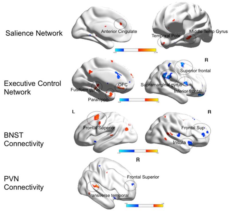
Objectives: Generalized anxiety disorder (GAD) is one of the most prevalent mental disorders in the elderly, but its functional neuroanatomy is not well understood. Given the role of emotion dysregulation in GAD, we sought to describe the neural bases of emotion regulation in late-life GAD by analyzing the functional connectivity (FC) in the Salience Network and the Executive Control Network during worry induction and worry reappraisal.

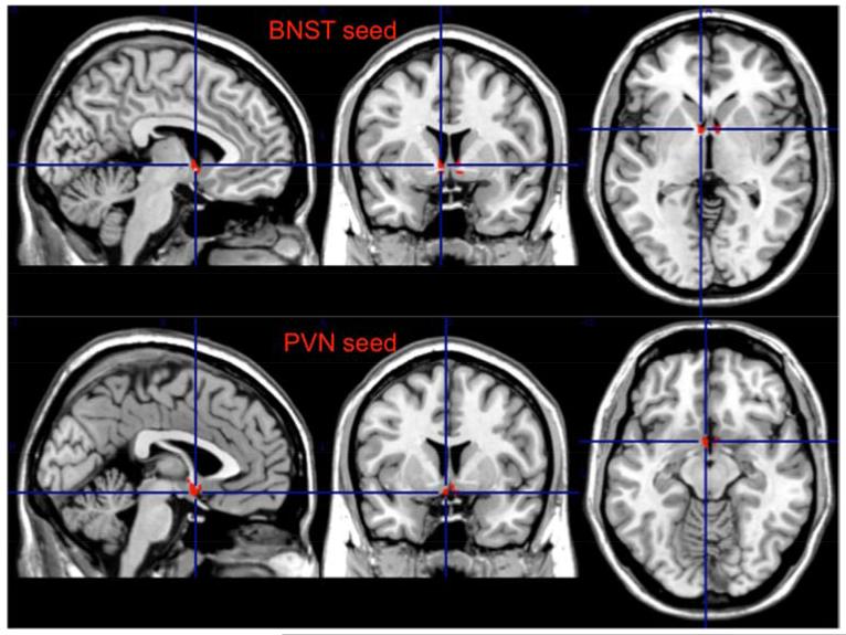
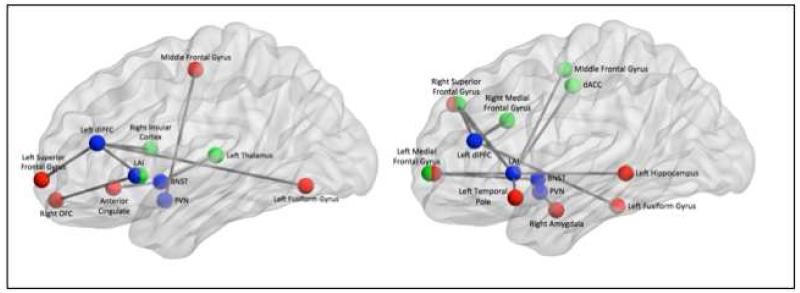
Methods: The study included 28 elderly GAD and 31 non-anxious comparison participants. Twelve elderly GAD completed a 12-week pharmacotherapy trial. We used an in-scanner worry script that alternates blocks of worry induction and reappraisal. We assessed network FC, using the following seeds: anterior insula (AI), dorsolateral prefrontal cortex (dlPFC), the bed nucleus of stria terminalis (BNST), and the paraventricular nucleus (PVN).

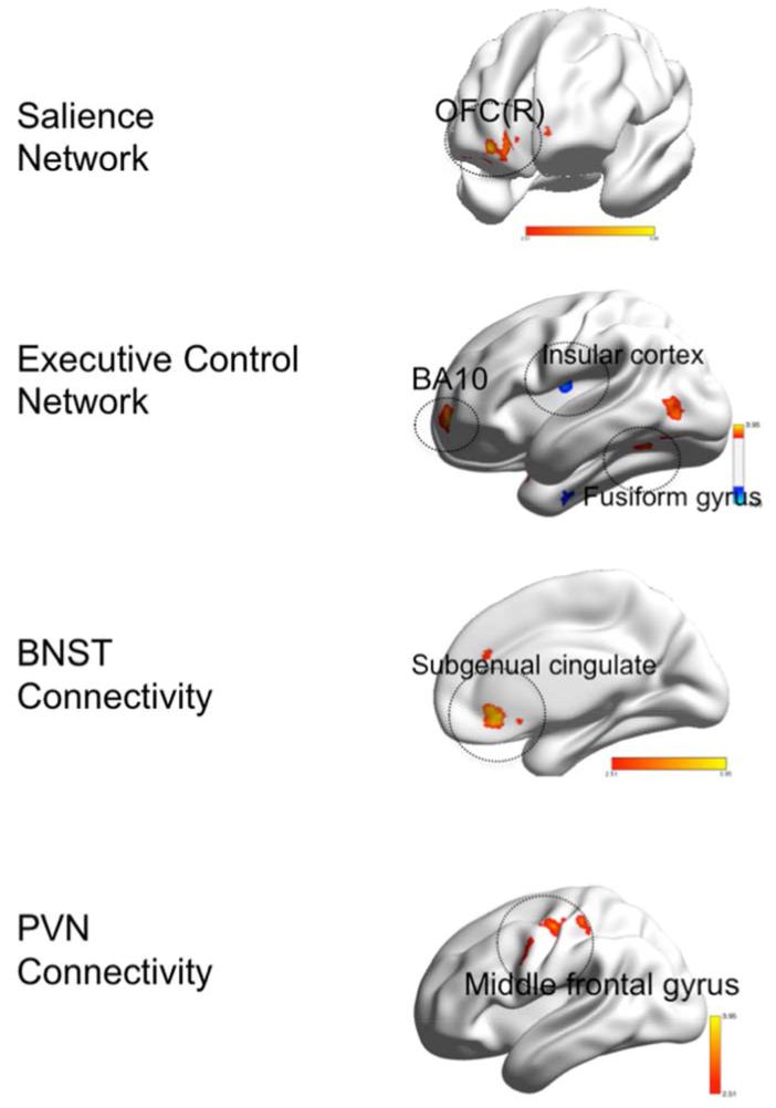
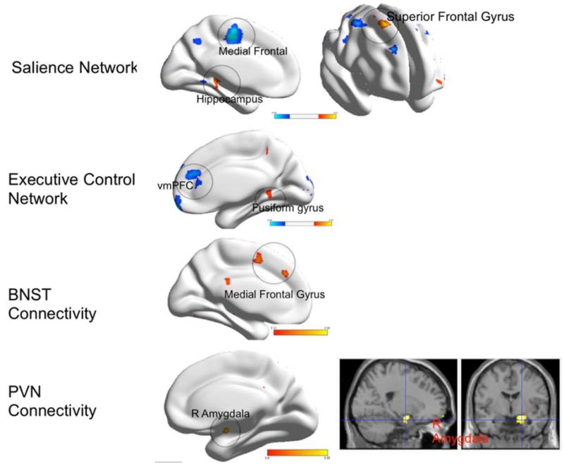
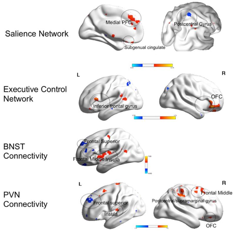
Results: GAD participants exhibited greater FC during worry induction between the left AI and the right orbitofrontal cortex, and between the BNST and the subgenual cingulate. During worry reappraisal, the non-anxious participants had greater FC between the left dlPFC and the medial PFC, as well as between the left AI and the medial PFC, and elderly GAD patients had greater FC between the PVN and the amygdala. Following 12 weeks of pharmacotherapy, GAD participants had greater connectivity between the dlPFC and several prefrontal regions during worry reappraisal.

Conclusion: FC during worry induction and reappraisal points toward abnormalities in both worry generation and worry reappraisal. Following successful pharmacologic treatment, we observed greater connectivity in the prefrontal nodes of the Executive Control Network during reappraisal of worry.

Keywords: Functional connectivity; emotion regulation; late-life generalized anxiety disorder.


Figures

Figure 1
Figure 1
The Bed Nucleus of Stria Terminalis (BNST) and Paraventricular Nucleus (PVN) seeds
Figure 2
Figure 2
Group differences in functional connectivity during worry induction between elderly GAD participants and elderly non-anxious participants Legend. Red: GAD>non-anxious comparison, blue: non-anxious comparison>GAD. BNST= bed nucleus of stria terminalis. PVN= paraventricular nucleus. OFC(R)=orbito-frontal gyrus, right. BA= Brodmann Area. Visualized using BrainNet Viewer, version 1.42 (64).Salience Network: OFC(R) (t=3.83; df=54). Executive Control Network: BA10 (t=3.71; df=54); Insular cortex (t=4.00; df=54); Fusiform gyrus (t-3.44; df=54). BNST Connectivity: Subgenual cingulate (t=3.95; df=54). PVN Connectivity: Middle frontal gyrus (t=4.25; df=54).
Figure 3
Figure 3
Group differences in functional connectivity during worry reappraisal between elderly GAD participants and non-anxious comparison participants Legend. Red: GAD>non-anxious comparison, blue: non-anxious comparison>GAD. BNST= bed nucleus of stria terminalis. PVN= paraventricular nucleus. vmPFC= ventromedial prefrontal cortex. BA= Brodmann Area. Visualized using BrainNet Viewer, version 1.42(64). Salience Network: Medial Frontal (t=4.26; df=54); Hippocampus (t=4.28; df=54); Superior frontal gyrus (t=4.44; df=54). Executive Control Network: vmPFC (t=4.41; df=54); Fusiform gyrus (t=4.59; df=54). BNST Connectivity: Medial frontal gyrus (t=4.08; df=54). PVN Connectivity: Amygdala(R) (t=4.88; df=54).
Figure 4
Figure 4
Within group differences in functional connectivity during worry induction between pre- and post-treatment elderly GAD participants Legend. Red: pre>post-treatment. Blue: post>pre-treatment. BNST= bed nucleus of stria terminalis. PVN= paraventricular nucleus. L=left, R=Right. OFC=orbito-frontal cortex. PFC=prefrontal cortex. Visualized using BrainNet Viewer, version 1.42(64). Salience Network: Medial PFC (t=6.0; df=11); Subgenual cingulate (t=5.43; df=11); Postcentral gyrus (t=5.1; df=11). Executive Control Network: Inferior frontal gyrus (t=7.3; df=11); OFC (t=6.7; df=11). BNST Connectivity: Frontal superior gyrus (t=5.6; df=11); Frontal middle (t=6.6; df=11); Insula (t=7.8; df=11). PVN Connectivity: Frontal superior (t=5.3; df=11); Insula (t=5.79, df=11); Postcentral/supramarginal gyrus (t=6.6; df=11); Frontal middle (t=5.4, df=11); OFC (t=6.4; df=11).
Figure 5
Figure 5
Within group differences in functional connectivity during worry reappraisal between pre- and post-treatment elderly GAD participants Legend. Red: pre>post-treatment. Blue: post>pre-treatment. BNST= bed nucleus of stria terminalis. PVN= paraventricular nucleus. L=left, R=Right. OFC=orbito-frontal cortex. Parahippo=parahippocampal cortex; PFC=prefrontal cortex. Visualized using BrainNet Viewer, version 1.42(64). Salience Network: Anterior cingulate (t=12.7; df=11); Temporal pole (t=7.4; df=11); Middle Temporal Gyrus (t=9.1; df=11). Executive Control Network: Fusiform gyrus (t=11.6; df=11); Parahippocampus (t=8.4; df=11); OFC (t=9.1; df=11); Superior frontal (t=7.2; df=11); Supramarginal gyrus (t=13.2, df=11); Inferior frontal (t=6.9; df=11). BNST Connectivity: Frontal superior (L) (t=7.4, df=11); Insula (t=8.9; df=11); Frontal superior (R) (t=5.4; df=11). PVN Connectivity: Transverse temporal (t=6.5; df=11); frontal superior (5.7; df=11).
Fig 6
Fig 6
Summarized findings showing differences in functional connectivity between non-anxious participants and elderly GAD during worry induction (left) and during worry reappraisal (right) Legend: In blue – the four seeds (LAI=left anterior insula, dlPFC=dorso-lateral prefrontal cortex, BNST=bed nucleus of stria terminalis, PVN=paraventricular nucleus. In red-regions of interest that had greater connectivity with the seed for GAD than for non-anxious participants. In green: regions of interest that had greater connectivity with the seed for non-anxious participants than for GAD.

References

    1. Beekman AT, Bremmer MA, Deeg DJ, et al. Anxiety disorders in later life: a report from the Longitudinal Aging Study Amsterdam. Int J Geriatr Psychiatry. 1998;13:717–726. - PubMed
    1. Le Roux H, Gatz M, Wetherell JL. Age at onset of generalized anxiety disorder in older adults. Am J Geriatr Psychiatry. 2005;13:23–30. - PubMed
    1. Flint AJ. Generalised anxiety disorder in elderly patients: epidemiology, diagnosis and treatment options. Drugs Aging. 2005;22:101–114. - PubMed
    1. de Beurs E, Beekman AT, van Balkom AJ, et al. Consequences of anxiety in older persons: its effect on disability, well-being and use of health services. Psychol Med. 1999;29:583–593. - PubMed
    1. Wetherell JLGM, Pedersen NL. A longitudinal analysis of anxiety and depressive symptoms. Psychol Aging. 2001;16:187–195. - PubMed

Publication types

MeSH terms