

Iterative reconstruction technique with reduced volume CT dose index: diagnostic accuracy in pediatric acute appendicitis
- PMID: 24996812
- PMCID: PMC4308578
- DOI: 10.1007/s00247-014-3109-7
Iterative reconstruction technique with reduced volume CT dose index: diagnostic accuracy in pediatric acute appendicitis
Abstract
Background: Iterative reconstruction technique has been proposed as a means of reducing patient radiation dose in pediatric CT. Yet, the effect of such reductions on diagnostic accuracy has not been thoroughly evaluated.
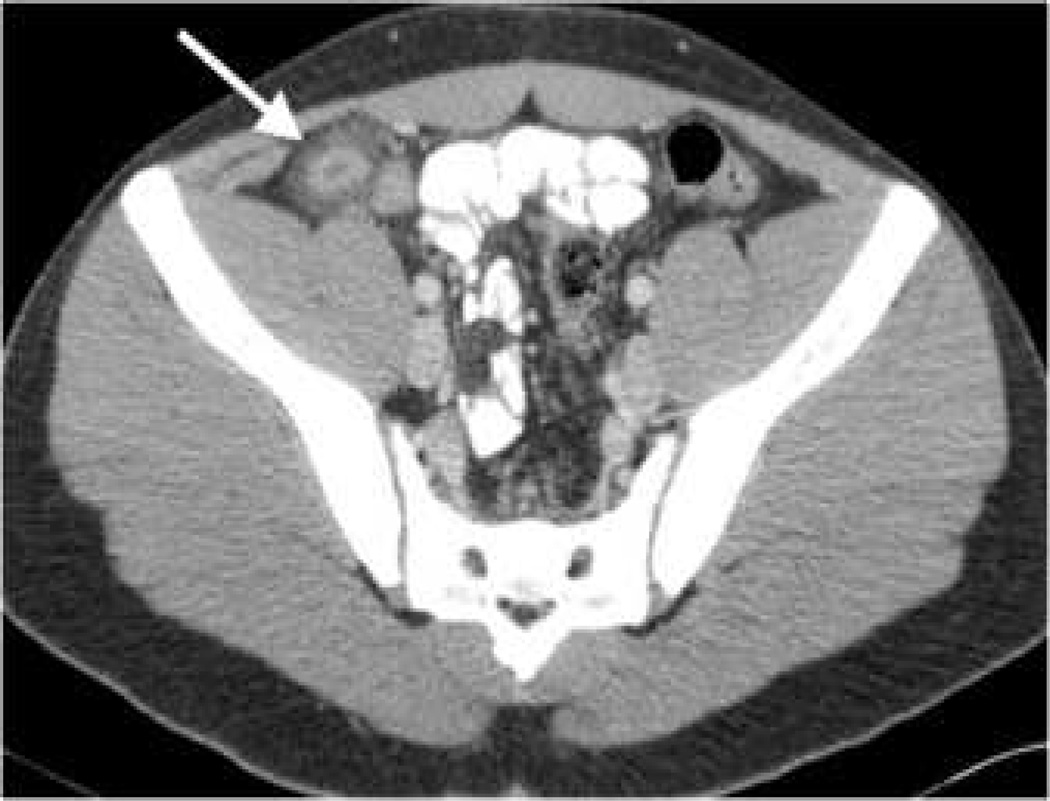
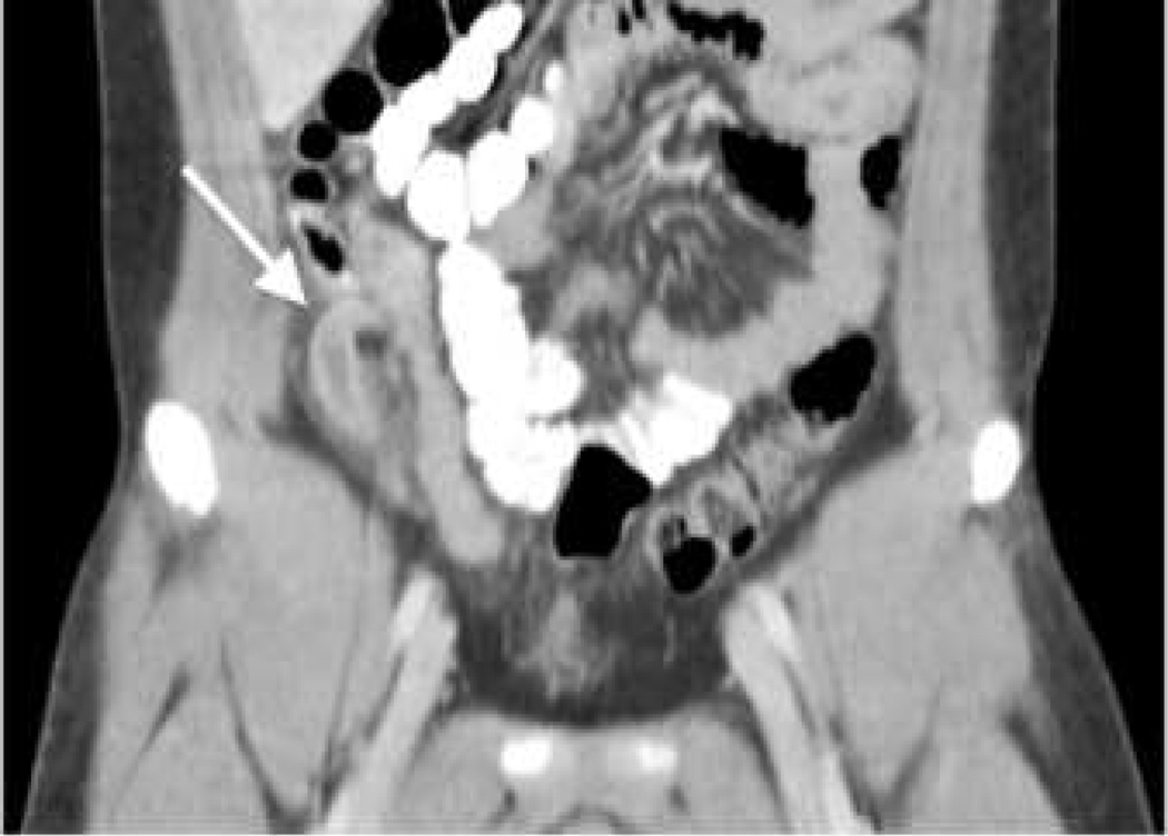
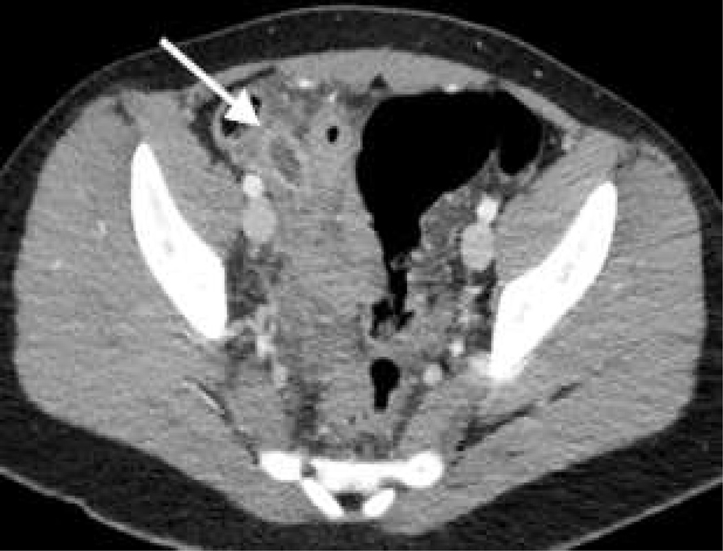
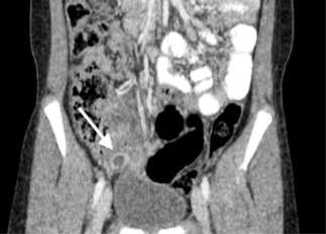
Objective: This study compares accuracy of diagnosing pediatric acute appendicitis using contrast-enhanced abdominopelvic CT scans performed with traditional pediatric weight-based protocols and filtered back projection reconstruction vs. a filtered back projection/iterative reconstruction technique blend with reduced volume CT dose index (CTDIvol).
Materials and methods: Results of pediatric contrast-enhanced abdominopelvic CT scans done for pain and/or suspected appendicitis were reviewed in two groups: A, 192 scans performed with the hospital's established weight-based CT protocols and filtered back projection reconstruction; B, 194 scans performed with iterative reconstruction technique and reduced CTDIvol. Reduced CTDIvol was achieved primarily by reductions in effective tube current-time product (mAseff) and tube peak kilovoltage (kVp). CT interpretation was correlated with clinical follow-up and/or surgical pathology. CTDIvol, size-specific dose estimates (SSDE) and performance characteristics of the two CT techniques were then compared.
Results: Between groups A and B, mean CTDIvol was reduced by 45%, and mean SSDE was reduced by 46%. Sensitivity, specificity and diagnostic accuracy were 96%, 97% and 96% in group A vs. 100%, 99% and 99% in group B.
Conclusion: Accuracy in diagnosing pediatric acute appendicitis was maintained in contrast-enhanced abdominopelvic CT scans that incorporated iterative reconstruction technique, despite reductions in mean CTDIvol and SSDE by nearly half as compared to the hospital's traditional weight-based protocols.
Conflict of interest statement
Figures
References
-
- Callahan MJ, Rodriguez DP, Taylor GA. CT of appendicitis in children. Radiology. 2002;224:325–332. - PubMed
-
- York D, Smith A, Phillips JD, von Allmen D. The influence of advanced radiographic imaging on the treatment of pediatric appendicitis. J Pediatr Surg. 2005;40:1908–1911. - PubMed
-
- Martin AE, Vollman D, Adler B, Caniano DA. CT scans may not reduce the negative appendectomy rate in children. J Pediatr Surg. 2004;39:886–890. discussion 890. - PubMed
-
- Doria AS, Moineddin R, Kellenberger CJ, et al. US or CT for diagnosis of appendicitis in children and adults? A meta-analysis. Radiology. 2006;241:83–94. - PubMed
-
- Taylor GA, Wesson DE. Acute appendicitis in children: diagnostic imaging. In: Singer JI, Wiley JF, editors. UpToDate®. Philadelphia, PA: Wolters Kluwer Health; 2013. [Accessed 18 December 2013]. http://www.uptodate.com.
Publication types
MeSH terms
Substances
Grants and funding
LinkOut - more resources
Full Text Sources
Other Literature Sources
Medical
