. 2014 Nov;24(11):1022-35.
doi: 10.1093/glycob/cwu064. Epub 2014 Jul 4.

Galectin-3 promotes HIV-1 budding via association with Alix and Gag p6

Affiliations

Galectin-3 promotes HIV-1 budding via association with Alix and Gag p6

Sheng-Fan Wang et al. Glycobiology. 2014 Nov.

Abstract

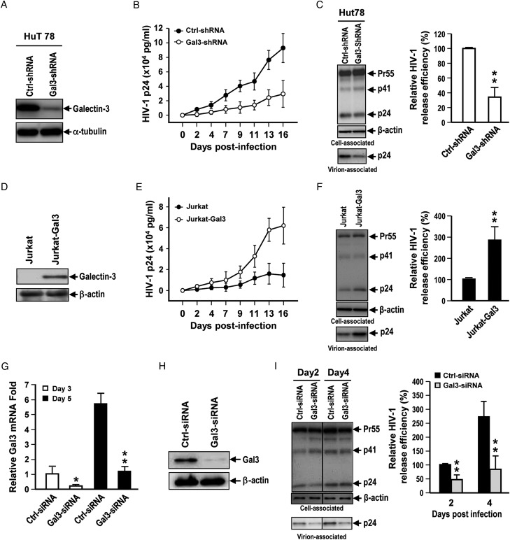
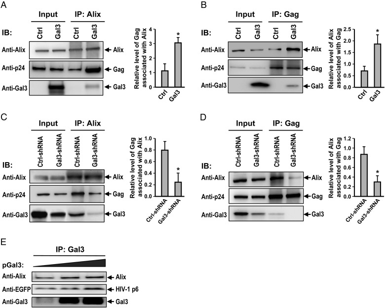
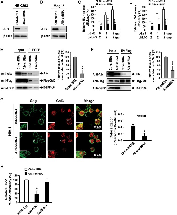
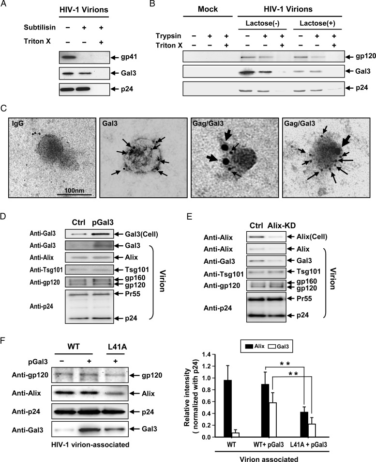
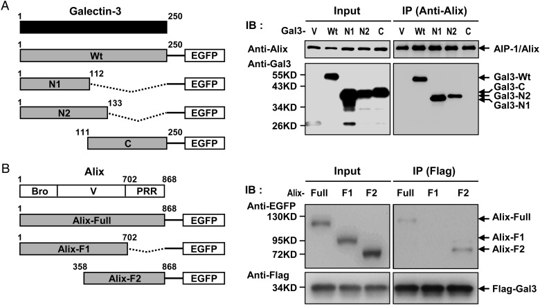
Galectin-3 has been reported to regulate the functions of a number of immune cell types. We previously reported that galectin-3 is translocated to immunological synapses in T cells upon T-cell receptor engagement, where it associates with ALG-2-interacting protein X (Alix). Alix is known to coordinate with the endosomal sorting complex required for transport (ESCRT) to promote human immunodeficiency virus (HIV)-1 virion release. We hypothesized that galectin-3 plays a role in HIV-1 viral budding. Cotransfection of cells of the Jurkat T line with galectin-3 and HIV-1 plasmids resulted in increased HIV-1 budding, and suppression of galectin-3 expression by RNAi in Hut78 and primary CD4+ T cells led to reduced HIV-1 budding. We used immunofluorescence microscopy to observe the partial colocalization of galectin-3, Alix and Gag in HIV-1-infected cells. Results from co-immunoprecipitation experiments indicate that galectin-3 expression promotes Alix-Gag p6 association, whereas the results of Alix knockdown suggest that galectin-3 promotes HIV-1 budding through Alix. HIV-1 particles released from galectin-3-expressing cells acquire the galectin-3 protein in an Alix-dependent manner, with proteins primarily residing inside the virions. We also found that the galectin-3 N-terminal domain interacts with the proline-rich region of Alix. Collectively, these results suggest that endogenous galectin-3 facilitates HIV-1 budding by promoting the Alix-Gag p6 association.

Keywords: Alix; HIV-1; galectin-3; viral budding.


Figures

Fig. 1.
Fig. 1.
Endogenous Galectin-3 enhances HIV-1 virus release. (A) Lentiviral shRNA-mediated knockdown of galectin-3 was performed in Hut78 cells and galectin-3 levels were determined by immunoblotting. (B) Control and galectin-3-knockdown Hut78 cells were infected with NL4-3 virions. Supernatants were collected at different time points for HIV-1 p24 measurement by enzyme-linked immunosorbent assay (ELISA). (C) Viral supernatants and cell lysates were collected for immunoblotting analysis and ELISA for HIV-1 p24 on day 2 postinfection. Relative HIV-1 release efficiency was calculated by dividing the amount of Gag(p24) in viral lysates by the total amount of Gag(p24) in cell and viral lysates. (D) Lentivirus-mediated galectin-3 expression was performed in Jurkat cells and galectin-3 levels were determined by immunoblotting. (E) Jurkat and Jurkat-Gal-3 cells were infected with NL4-3 viruses. Supernatants were collected at different time points for HIV-1 p24 measurement by ELISA. (F) Viral supernatants and cell lysates were collected for immunoblotting analysis and ELISA for HIV-1 p24 on day 2 postinfection. Relative HIV-1 release efficiency was calculated in (C). (G) Human primary CD4+ T cells were subjected to galectin-3 knockdown by treatment with siRNAs. Relative mRNA levels of galectin-3 in control and galectin-3-siRNA-treated primary CD4+ T cells cultured for 3 or 5 days were analyzed by quantitative RT-PCR. (H) Galectin-3 protein expression in control and galectin-3-siRNA-treated primary CD4+ T cells was analyzed by immunoblotting. (I) Control and galectin-3-siRNA-treated primary CD4+ T cells were infected with HIV-1; supernatants and cell lysates were collected for immunoblotting analysis and ELISA for HIV-1 p24, and relative HIV-1 release efficiency was calculated in (C). Quantitative data represent the means ± SD of results from three independent experiments. Significance values were calculated using two-tailed Student's t-tests (*P < 0.05; **P < 0.01).
Fig. 2.
Fig. 2.
Galectin-3 association with Alix in HIV-1-infected cells. (A) pFlag-Gal3 and pNL4-3 vectors were cotransfected into HEK293T cells, followed by incubation for 48 h at 37°C. After three washes with PBS containing 30 mM lactose, cells were treated with the chemical cross-linker DSP, lysed and immunoprecipitated with rabbit anti-Flag (left and right panel) or rabbit anti-Alix (middle panel) antibodies. Precipitated proteins were analyzed by immunoblotting for Flag, galectin-3, Alix and Tsg101 proteins. H-chain indicates IgG heavy chain; C and T refer to control (nontransfected) and transfected groups, respectively. Magi-5 (B) and primary human CD4+ cells (C) were infected with HIV-1 NL4-3 viruses. After 24 h, cells were fixed and stained with antibodies against the indicated proteins and observed using confocal microscopy. Representative cells are shown. Colocalization values for Gag (green) with Alix (red) and for Alix (red) with galectin-3 (blue) in 50 stained cells of each type were measured using Global Pearson's Correlation Coefficient statistics, in Volocity version 6.1.1 (PerkinElmer). (D) HIV-1-infected Jurkat-Gal3T cells were fixed and stained with antibodies against the indicated proteins. Stained cells were analyzed by TIRF and SR fluorescence microscopy. The images were recorded from the lower membrane of the cells and each dot in the SR images represents a protein molecule. The colocalization coefficients of two proteins on the lower membranes of single cells were determined by Manders' analysis (Manders et al. 1993). Protein colocalization was quantified using two-dimensional spatial histogram maps of two fluorescence channels, with fluorescence backgrounds removed during the intensity-based colocalization analyses. Results from one representative experiment of three independently performed experiments are shown.
Fig. 3.
Fig. 3.
Galectin-3 promotes Alix-Gag association. HEK293T cells were cotransfected with pNL4-3, pEF1-Gal3 or a control pEF-1 vector, lysed, immunoprecipitated with anti-Alix (A) or anti-Gag (B) antibodies, and immunoblotted with the indicated antibodies. Immunoblot band intensities were quantified by densitometry. Amounts of co-immunoprecipitated Gag (A) or Alix (B) were normalized to pulled down Alix (A) or Gag (B) levels, subsequently. (C) Control and galectin-3-knockdown Magi-5 cells were transfected with pNL4-3, lysed, immunoprecipitated with anti-Alix (C) or anti-Gag (D) antibodies, and subjected to immunoblotting with the indicated antibodies. Immunoblot band intensities were quantified by densitometry. Amounts of co-immunoprecipitated Gag (C) or Alix (D) were subsequently normalized to pulled down Alix levels (C) or Gag (D) levels. (E) pEGFP-p6 (2 μg) was cotransfected with different concentrations of pEF1-Gal3 (0–2 μg) into HEK293T cells. Cell lysates were subjected to co-immunoprecipitation assays with anti-galectin-3, followed by immunoblotting with the indicated antibodies. Representative immunoblotting results are shown. Quantitative data represent the means ± SD of results from three independent experiments. Significance values were calculated using two-tailed Student's t-tests (*P < 0.05; **P < 0.01).
Fig. 4.
Fig. 4.
Galectin-3 depends on Alix to facilitate HIV-1 budding. (A and B) Lentivirus shRNA-mediated Alix knockdown in HEK293T and Magi-5 cells was analyzed by immunoblotting. (C and D) Control and Alix-knockdown HEK293T and Magi-5 cells were cotransfected with different ratios of pNL4-3 and pEF1-Gal3. Supernatants and cell lysates were collected for HIV-1 p24 ELISA. Relative HIV-1 release efficiency was calculated by dividing the amount of Gag(p24) in viral lysates by the total amount of Gag(p24) in cell and viral lysates. (E) Control and Alix-knockdown HEK293T cells were cotransfected with pEGFP-p6 and pFlag-Gal3. The lysates were subjected to a co-immunoprecipitation assay followed by immunoblot analysis; the band intensities were quantified by densitometry. Amounts of co-immunoprecipitated galectin-3 were normalized to the levels of pulled down p6, and the normalized quantities of galectin-3 for Alix-knockdown cells relative to the control group are shown. (F) Control and Alix-knockdown HEK293T cells were cotransfected with pEGFP-p6 and pFlag-Gal3. The lysates were subjected to a co-immunoprecipitation assay followed by immunoblot analysis; the band intensities were quantified by densitometry. Amounts of co-immunoprecipitated p6 were normalized to the pulled down galectin-3 levels and the normalized quantities of p6 for Alix-knockdown cells relative to the control group are shown. (G) HIV-1 infected control and Alix-knockdown Hut78 cells were stained with antibodies against HIV-1 Gag and galectin-3 prior to observation using confocal microscopy. Colocalization values for Gag (green) with galectin-3 (red) in 100 control or Alix-knockdown cells were measured using Global Pearson's Correlation Coefficient statistics. (H) pEGFP (EGFP-Ctrl) or pEGFP-Alix (EGFP-Alix) was transfected into control and galectin-3 knockdown Hut78T cells. Following HIV-1 infection, supernatants and cell lysates were collected for p24 ELISAs, and relative HIV-1 release efficiency was calculated by dividing the amount of Gag(p24) in viral lysates by the total amount of Gag(p24) in cell and viral lysates. In Quantitative data represent the means ± SD of results from three independent experiments. Significance values were calculated using two-tailed Student's t-tests (*P < 0.05; **P < 0.01).
Fig. 5.
Fig. 5.
Presence of Galectin-3 in HIV-1 virions. pEF1-Gal3 and pNL4-3 plasmids were cotransfected into HEK293T cells. Supernatants were collected for HIV-1 virion purification via sucrose gradients. (A) Sucrose gradient-purified HIV-1 virions (100 ng, as quantified by p24 ELISAs) were left untreated or were treated with subtilisin (1 mg/mL) at room temperature for 1 h in the absence or presence of Triton X-100 (1.5%). Treated and untreated virions were subjected to immunoblotting analyses for each indicated protein. (B) Purified HIV-1 virions (100 ng, as quantified by p24 ELISAs) were left untreated or were treated with lactose and subjected to trypsin digestion (150 μg/mL) at room temperature for 1 h in the absence or presence of Triton X-100 (1.5%). Treated and untreated virions were subjected to immunoblotting analyses for each indicated protein. (C) Purified virions were loaded on grids, fixed and stained with goat anti-galectin-3 (Gal3), mouse anti-p24 (Gag) or control IgG (IgG), followed by Donkey anti-goat-labeled 6-nm gold particles and Donkey anti-mouse-labeled 18-nm gold particles. Following 4% uranyl acetate staining, grids were examined by TEM (scale bar =100 nm; thin and thick arrows indicate galectin-3 and Gag protein localization, respectively). (D) Purified HIV-1 virions were analyzed by immunoblotting for each indicated protein. (E) Alix-knockdown Magi-5 cells were transfected with pNL4-3 and incubated at 37°C for 48 h. The supernatants were collected for HIV-1 virion purification. Purified virions and cell lysates were subjected to immunoblotting assays for the indicated proteins. (F) HEK293T cells were transfected with pNL4-3 or pL41A together with pGal3. The supernatants were collected for HIV-1 virion purification. Purified HIV-1 wild-type or L41A mutant virions were analyzed by immunoblotting for each indicated protein.
Fig. 6.
Fig. 6.
(A) Galectin-3 N-terminal region interacts with the PRR of Alix. Various truncated forms of galectin-3 (wild type, N1, N2 and C domains) were cloned into pEFGP-N1 and used to cotransfect HEK293T cells with pNL4-3. Transfected cell lysates were immunoprecipitated with anti-Alix and precipitated proteins were subjected to immunoblotting with anti-galectin-3 antibodies. V, vector control; Wt, wild type; N1, N-terminal region (residues 1–112) of galectin-3; N2, N-terminal region (residues 1–133) of galectin-3; C, C-terminal region containing the entire CBD domain (residues 111–250) of galectin-3. (B) Various truncated forms of Alix (including Full, F1 and F2) were cloned into pEFGP-N1 and used to cotransfect HEK293T cells with pFlag-Gal3 and pNL4-3. Cell lysates were immunoprecipitated with antibodies against Flag; precipitated proteins were immunoblotted with anti-Alix antibodies.

Similar articles

Cited by

References

    1. Booth AM, Fang Y, Fallon JK, Yang JM, Hildreth JE, Gould SJ. Exosomes and HIV Gag bud from endosome-like domains of the T cell plasma membrane. J Cell Biol. 2006;172:923–935. - PMC - PubMed
    1. Caballe A, Martin-Serrano J. ESCRT machinery and cytokinesis: The road to daughter cell separation. Traffic. 2011;12:1318–1326. - PubMed
    1. Carlton JG, Agromayor M, Martin-Serrano J. Differential requirements for Alix and ESCRT-III in cytokinesis and HIV-1 release. Proc Natl Acad Sci USA. 2008;105:10541–10546. - PMC - PubMed
    1. Chen HY, Fermin A, Vardhana S, Weng IC, Lo KF, Chang EY, Maverakis E, Yang RY, Hsu DK, Dustin ML, et al. Galectin-3 negatively regulates TCR-mediated CD4+ T-cell activation at the immunological synapse. Proc Natl Acad Sci USA. 2009;106:14496–14501. - PMC - PubMed
    1. Chen C, Vincent O, Jin J, Weisz OA, Montelaro RC. Functions of early (AP-2) and late (AIP1/ALIX) endocytic proteins in equine infectious anemia virus budding. J Biol Chem. 2005;280:40474–40480. - PubMed

Publication types

MeSH terms

Substances