. 2014 Jul 4:14:483.
doi: 10.1186/1471-2407-14-483.

γH2AX and Chk1 phosphorylation as predictive pharmacodynamic biomarkers of Chk1 inhibitor-chemotherapy combination treatments

Affiliations

γH2AX and Chk1 phosphorylation as predictive pharmacodynamic biomarkers of Chk1 inhibitor-chemotherapy combination treatments

Rebecca Rawlinson et al. BMC Cancer. .

Abstract

Background: Chk1 inhibitors are currently in clinical trials in combination with a range of cytotoxic agents and have the potential to potentiate the clinical activity of a large number of standard of care chemotherapeutic agents. Utilizing pharmacodynamic biomarkers to optimize drug dose and scheduling in these trials could greatly enhance the likelihood of clinical success.

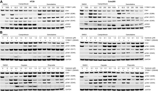
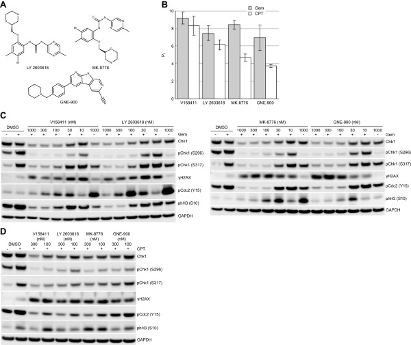
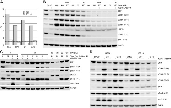
Methods: In this study, we evaluated the in vitro potentiation of the cytotoxicity of a range of cytotoxic chemotherapeutic drugs by the novel Chk1 inhibitor V158411 in p53 mutant colon cancer cells. Pharmacodynamic biomarkers were evaluated in vitro.

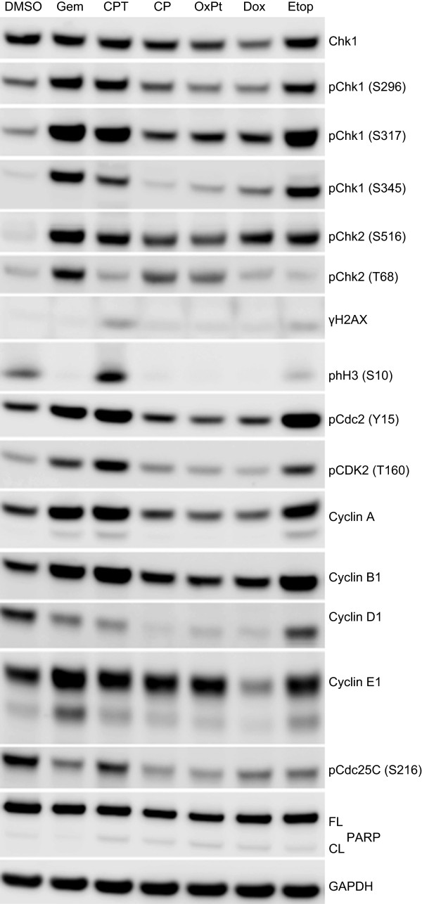
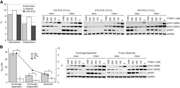
Results: V158411 potentiated the cytotoxicity of a range of chemotherapeutic agents with distinct mechanisms of action in p53 mutant colon cancer cell lines grown in anchorage dependent or independent culture conditions. Analysis of pharmacodynamic biomarker changes identified dependencies on the chemotherapeutic agent, the concentration of the chemotherapeutic and the duration of time between combination treatment and biomarker analysis. A reduction in total Chk1 and S296/S317/S345 phosphorylation occurred consistently with all cytotoxics in combination with V158411 but did not predict cell line potentiation. Induction of γH2AX levels was chemotherapeutic dependent and correlated closely with potentiation of gemcitabine and camptothecin in p53 mutant colon cancer cells.

Conclusions: Our results suggest that Chk1 phosphorylation could be a useful biomarker for monitoring inhibition of Chk1 activity in clinical trials involving a range of V158411-chemotherapy combinations and γH2AX induction as a predictor of potentiation in combinations containing gemcitabine or camptothecin.


Figures

Figure 1
Figure 1
Determination of in vitro potentiation of cytotoxic chemotherapeutic agents by V158411. A. Comparison of potentiation factors in HT29 and Colo205 cancer cells grown anchorage dependently, anchorage independently or as multicellular tumor spheroids for cytotoxic chemotherapeutic agents with 400 nM V158411. Pf values were calculated from the average GI50 and combination GI50 (cGI50) of at least three determinations where Pf equals average GI50/average cGI50. *, P < 0.05; **, P < 0.01; ND, not determinable; #, Pf > 15. B. 72 hour HT29 dose response curves for gemcitabine, camptothecin and cisplatin in combination with DMSO or 400 nM V158411 demonstrating the calculation of the GI50 (square and dotted line) and cGI50 (circle and line). C. HT29 dose response curves for gemcitabine following 168 hour treatment with DMSO or 400 nM V158411. HT29 cells were grown either anchorage dependently (left graph) or as multi-cellular tumor spheroids (right graph). D. Comparison of the single agent GI50 and consequent potentiation factor (Pf, triangle and line) following either 72 hour (3 day) or 168 hour (7 day) incubation with gemcitabine, camptothecin or cisplatin with 0 or 400 nM V158411 in HT29 cells growing anchorage dependently. Values are the average of at least 3 determinations ± SD.
Figure 2
Figure 2
Checkpoint activation and DNA damage protein biomarker responses in HT29 cells following treatment with cytotoxic chemotherapeutic agents. HT29 cells were treated with approximately 5-times the single agent GI50 of gemcitabine (Gem, 0.2 μM), camptothecin (CPT, 1 μM), cisplatin (CP, 125 μM), oxaliplatin (OxPt, 250 μM), doxorubicin (Dox, 3 μM) or etoposide (Etop, 50 μM) for 24 hours. Changes in protein expression levels were determined by immunoblotting.
Figure 3
Figure 3
V158411 inhibits DNA damage induced Chk1 auto-phosphorylation and increases γH2AX in colon carcinoma cells. HT29 (left) or Colo205 (right) p53 defective colon carcinoma cells were treated with A. 200 nM camptothecin or 100 nM gemcitabine plus varying concentrations of V158411 for 24 hours or B. treated with the indicated concentrations of gemcitabine, camptothecin, cisplatin or etoposide with either DMSO or 200 nM V158411 for 24 hours. Protein expression was characterized by immunoblotting.
Figure 4
Figure 4
Pharmacodynamic changes to DNA damage checkpoint and cell cycle proteins in HT29 colon cancer cells by V158411 in combination with camptothecin. A. HT29 cells were treated with 0 to 400 nM camptothecin (CPT) in the presence of DMSO or 400 nM V158411 for 24 hours. B. HT29 cells were treated with 100 nM camptothecin plus DMSO or 400 nM V158411 for 1 to 24 hours. UT, untreated control. C. HT29 cells were treated with 200 nM camptothecin for 0 to 24 hours followed by 400 nM V158411 (+) or DMSO (−) for a further 24 hours. Protein expression was characterized by immunoblotting.
Figure 5
Figure 5
Cellular biomarker responses in HT29 cells exposed to various cytotoxic chemotherapeutic agents in combination with the Chk1 inhibitor V158411. HT29 cells were exposed to the combination GI80 of gemcitabine (0.2 μM), camptothecin (0.44 μM), cisplatin (68 μM), oxaliplatin (131 μM), doxorubicin (1.2 μM) or etoposide (59 μM) for 18 hours followed by DMSO (−) or 400 nM V158411 (+) for a further 24 hours. Protein expression was characterized by immunoblotting.
Figure 6
Figure 6
Potentiation of gemcitabine and camptothecin cytotoxicity and protein biomarker changes induced in HT29 cells by structurally diverse Chk1 inhibitors. A. Chemical structures of Chk1 inhibitors. B. Potentiation of gemcitabine (Gem) or camptothecin (CPT) cytotoxicity in HT29 cells following 72 hour exposure in combination with 300 nM V158411, LY2603618, MK-8776 or GNE-900. The potentiation factor (Pf) was calculated as GI50 cytotoxic agent alone/GI50 cytotoxic agent plus Chk1 inhibitor. Values represent the average of 3 determinations ± SD. C. Biomarker changes induced in response to gemcitabine plus Chk1 inhibitor treatment in HT29 colon carcinoma cells. HT29 colon cancer cells were exposed to 50 nM gemcitabine (+) for 16 hours followed by increasing concentrations of Chk1 inhibitor for a further 24 hours. Protein expression was characterized by immunoblotting. D. Biomarker changes induced in response to camptothecin plus Chk1 inhibitor treatment in HT29 colon carcinoma cells. HT29 colon cancer cells were exposed to 100 nM camptothecin for 16 hours followed by 100 or 300 nM Chk1 inhibitor for a further 24 hours.
Figure 7
Figure 7
Comparison of protein biomarker changes in p53 proficient and deficient colon cancer cell lines. A. Potentiation of the cytotoxicity of gemcitabine (Gem), camptothecin (CPT), cisplatin (CP) or oxaliplatin (OxPt) by 400 nM V158411 was determined in p53 mutant HT29 or p53 wild-type HCT116 colon cancer cells after 72 hours. GI50 and cGI50 were calculated from the dose response curves using XLFit. The potentiation factor (Pf) was calculated as GI50/cGI50. Protein biomarker changes were assessed in HCT116 cells treated with B. 50 to 800 nM gemcitabine or 120 nM camptothecin in combination with 0 (−) or 400 nM (+) V158411 for 24 hours or C. camptothecin plus 0 (−) or 400 nM (+) V158411 for various time combinations and dosing regimens. D. HT29 or HCT116 cells were treated with the single agent IC80 of camptothecin (HT29, 0.43 μM; HCT116, 0.44 μM) or oxaliplatin (HT29, 131 μM; HCT116, 74 μM) for 18 hours followed by DMSO (−) or 400 nM (+) V158411 for a further 24 hours. Protein expression was characterized by immunoblotting.
Figure 8
Figure 8
Potentiation of gemcitabine and camptothecin cytotoxicity by V158411 occurs independently of fetal calf serum or oxygen concentration and under anchorage independent growth conditions. A. Potentiation of the cytotoxicity of gemcitabine or camptothecin by 400 nM V158411 was determined in HT29 cells growing in 10% FCS, 21% O2 (normoxia); 10% FCS, 0.5% O2 (hypoxia) or 0.5% FCS, 21% O2 for 72 hours. GI50 and cGI50 were calculated from the dose response curves using XLFit. The potentiation factor (Pf) was calculated as the average GI50/average cGI50 from 3 determinations. Protein biomarker changes were subsequently assessed in HT29 cells growing under the same conditions following treatment with 100 nM gemcitabine plus 0 to 1000 nM V158411 for 24 hours. B. HT29 cells growing either attached to plastic cell culture plates (anchorage dependently), anchorage independently in LMP agarose or as multicellular tumor spheroids were exposed to increasing concentrations of gemcitabine in the presence or absence of 400 nM V158411 for 72 (anchorage dependent) or 168 (anchorage independent/spheroid) hours. **, P < 0.01. Protein biomarker changes were assessed in HT29 cells growing anchorage dependently or as multi-cellular tumor spheroids following treatment with 100 nM gemcitabine plus 0 to 1000 nM V158411 for 24 hours. Protein expression was characterized by immunoblotting.

References

    1. Liu Q, Guntuku S, Cui XS, Matsuoka S, Cortez D, Tamai K, Luo G, Carattini-Rivera S, DeMayo F, Bradley A, Donehower LA, Elledge SJ. Chk1 is an essential kinase that is regulated by Atr and required for the G(2)/M DNA damage checkpoint. Genes Dev. 2000;14:1448–1459. - PMC - PubMed
    1. Dai Y, Grant S. New insights into checkpoint kinase 1 in the DNA damage response signaling network. Clin Cancer Res. 2010;16:376–383. - PMC - PubMed
    1. Smith J, Tho LM, Xu N, Gillespie DA. The ATM-Chk2 and ATR-Chk1 pathways in DNA damage signaling and cancer. Adv Cancer Res. 2010;108:73–112. - PubMed
    1. Bucher N, Britten CD. G2 checkpoint abrogation and checkpoint kinase-1 targeting in the treatment of cancer. Br J Cancer. 2008;98:523–528. - PMC - PubMed
    1. Tapia-Alveal C, Calonge TM, O’Connell MJ. Regulation of chk1. Cell Div. 2009;4:8. - PMC - PubMed

Publication types

MeSH terms