Review
. 2014 Dec 26:283:78-94.
doi: 10.1016/j.neuroscience.2014.06.056. Epub 2014 Jul 2.

Cortical reorganization after spinal cord injury: always for good?

Affiliations
Review

Cortical reorganization after spinal cord injury: always for good?

K A Moxon et al. Neuroscience. .

Abstract

Plasticity constitutes the basis of behavioral changes as a result of experience. It refers to neural network shaping and re-shaping at the global level and to synaptic contacts remodeling at the local level, either during learning or memory encoding, or as a result of acute or chronic pathological conditions. 'Plastic' brain reorganization after central nervous system lesions has a pivotal role in the recovery and rehabilitation of sensory and motor dysfunction, but can also be "maladaptive". Moreover, it is clear that brain reorganization is not a "static" phenomenon but rather a very dynamic process. Spinal cord injury immediately initiates a change in brain state and starts cortical reorganization. In the long term, the impact of injury - with or without accompanying therapy - on the brain is a complex balance between supraspinal reorganization and spinal recovery. The degree of cortical reorganization after spinal cord injury is highly variable, and can range from no reorganization (i.e. "silencing") to massive cortical remapping. This variability critically depends on the species, the age of the animal when the injury occurs, the time after the injury has occurred, and the behavioral activity and possible therapy regimes after the injury. We will briefly discuss these dependencies, trying to highlight their translational value. Overall, it is not only necessary to better understand how the brain can reorganize after injury with or without therapy, it is also necessary to clarify when and why brain reorganization can be either "good" or "bad" in terms of its clinical consequences. This information is critical in order to develop and optimize cost-effective therapies to maximize functional recovery while minimizing maladaptive states after spinal cord injury.

Keywords: brain plasticity; brain-derived neurotrophic factor; exercise; pain; serotonin; spinal transection.


Figures

Figure 1
Figure 1
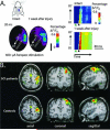
Expansion of the intact cortex into the deafferented cortex after spinal cord injury occurs in several species from rat to human. A. Reorganization in the rat hindlimb cortex after bilateral, dorsal hemisection as measured by voltage sensitive dye (VSD) imagining. Left top left: purple region in the schematic representation of the rat head shows the region of the brain where VSD imaging was performed. Left bottom: seven regions of interest (ROIs), 300-μm in diameter, were defined (R1–R7 white circles). R1 was placed adjacent to the forelimb representation and subsequent ROIs were placed progressively caudo-medial at an angle of 35° from the midline. Right: activation of the voltage sensitive dye across the ROIs as a function of time in response to electrical stimulation of the forepaw. Activation begins nearest the forelimb representation and is greater one week after injury. Green arrowheads denote the time of stimulation. Black arrowhead denotes 20 ms after stimulation. Figure reproduced from Ghosh et al., (2010) with permission. B. Reorganization in the human cortex after spinal cord injury as measured by functional magnetic resonance imagining (fMRI). These data represent group results from nine paraplegic patients approximately 40 months after injury to the thoracic or lumbar region. Subjects were asked to repetitively perform finger-to-thumb opposition of the digits 2, 3, 4 and 5. Images show group averages. The activation patterns in the SCI patients (top row) is significantly enlarged compared to that of the controls (bottom row) with a medial and lateral expansion of the volume and an additional increase in activation in the contralateral premotor and parietal areas. Figure reproduced from Curt et al. (2002), with permission.
Figure 2
Figure 2
The extent of reorganization after spinal cord injury is dependent on the time after injury. A. In primates, after unilateral, cervical dorsal column lesions, neurons in the deafferented cortex were unresponsive to sensory stimulation immediately after the lesion (95-12 acute, postsection). After 5 days, there was a significant enlargement of the intact regions above the level of the lesion (e.g. face) that then constricted by one month post-lesion. However, by 8 months post lesion, cells across the entire area 3b were responsive only to stimulation above the level of the lesion or the face (95-87 8 months). Figure reproduced from Jain et al. (1997), with permission. B. Similar results were found in the rat using fMRI. Group analysis of the responsiveness of cortex to electrical stimulation of the forepaw in 12 rats transected at T9. Coronal sections are along the top and horizontal sections are along the bottom. Significant enlargement of the fMRI signal, extending both medially and caudally (into the hindlimb cortex) from the original forelimb begins 3 days after injury and is then lost approximately 1-2 weeks after injury. By 1-2 months after injury, the expansion is reestablished and continues to enlarge by 3-6 months. Figure reproduced from Endo et al. (2007), with permission. C. Further studies in the rat show that urethane-induced delta oscillations (1-4Hz) before spinal transection (left panels showing local field potential, LFP, and multiunit activity, MUA) transitioned to slow-wave oscillations (<1Hz) immediately (i.e. within minutes) after spinal transection (middle panels), corresponding to a reduction in spontaneous activity as evidenced by a decrease mean amplitude of the rectified multi-unit activity (rMUA) and a decreased frequency of the rMUA spectrum (right panel). Figure reproduced from Aguilar et al. (2010), with permission.
Figure 3
Figure 3
Serotonin is known to promote adult cortical plasticity after injury. A. Studies have shown that pharmacotherapy after injury can increase BNDF-trkB signaling which increases the remodeling of chromatin structures ultimately lowering the threshold for cortical excitability, thus increasing the probability for sensory stimulation (in this case visual) to drive activity-dependent modification of synaptic transmission. Figure reproduced from Maya-Vetencourt et al. (2011), with permission. B. Similar results were observed after spinal cord injury. Top panel shows the responsiveness of the hindlimb sensory-motor cortex (HLSM, oval outlined in black) and forelimb motor cortex (quadrilateral located more rostrally) to tactile stimulation of the forepaw. Responsiveness is measured by the number of neurons that respond to the forepaw stimulation with increased firing rate. Middle panel shows expansion of the response into both the hindpaw sensory cortex and forepaw motor cortex after 8-weeks of a daily injection of a low dose (LD) of 5-HT agonists. Bottom panel shows greater expansion after 8-weeks of daily injection of a high dose (HD) of 5-HT agonists. Responses in the forepaw cortex (black outline more lateral from the hindpaw are not shown). Figure reproduced from Ganzer et al. (2013), with permission.
Figure 4
Figure 4
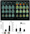
Exercise promotes cortical reorganization after spinal cord injury. A. Robotic locomotor therapy that improved functional outcome also enhanced motor representation of the foot. Images are an example from one patient. The red-orange areas show regions of brain activation in response to voluntary ankle plantar flexion and toe flection as measure by fMRI. Figure reproduced from Winchester et al. (2005), with permission. B. Similar results were found for spinalized rats after 8-weeks of passive bike exercise below the level of the lesion. The exercise increased the levels of adenylate cyclase 1 (ADY1) and brain-derived neurotrophic factor (BDNF) in the cortex. C. Bike exercise also increased the probability that cells in the deafferented hindlimb cortex would respond to tactile stimulation of the forelimbs across all cortical layers: supragranular (SP), granular (G) and infragranular (IG). Figure reproduced from Graziano et al. (2013), with permission.

References

    1. Aguilar J, Humanes-Valera D, Alonso-Calvino E, Yague JG, Moxon KA, Oliviero A, Foffani G. Spinal cord injury immediately changes the state of the brain. J Neurosci. 2010;30:7528–7537. - PMC - PubMed
    1. Aguilar JR, Castro-Alamancos MA. Spatiotemporal gating of sensory inputs in thalamus during quiescent and activated states. J Neurosci. 2005;25:10990–11002. - PMC - PubMed
    1. Angeli CA, Edgerton VR, Gerasimenko YP, Harkema SJ. Altering spinal cord excitability enables voluntary movements after chronic complete paralysis in humans. Brain. 2014;137:1394–1409. - PMC - PubMed
    1. Antri M, Barthe JY, Mouffle C, Orsal D. Long-lasting recovery of locomotor function in chronic spinal rat following chronic combined pharmacological stimulation of serotonergic receptors with 8-OHDPAT and quipazine. Neurosci Lett. 2005;384:162–167. - PubMed
    1. Antri M, Mouffle C, Orsal D, Barthe JY. 5-HT1A receptors are involved in short- and long-term processes responsible for 5-HT-induced locomotor function recovery in chronic spinal rat. Eur J Neurosci. 2003;18:1963–1972. - PubMed

Publication types