. 2015 Oct;67(5):851-60.
doi: 10.1007/s10616-014-9725-0. Epub 2014 Jul 6.

Establishment of a xeno-free culture system that preserves the characteristics of placenta mesenchymal stem cells

Affiliations

Establishment of a xeno-free culture system that preserves the characteristics of placenta mesenchymal stem cells

Saeyoung Park et al. Cytotechnology. 2015 Oct.

Abstract

Although stem cells are promising candidates for cell replacement therapies, the vast majority are derived using animal sera, which has risk of being contaminated by animal viruses or toxins. To overcome these potential problems, we initially established multiple lines of stem cells from first-trimester human placenta (fPMSC), which were cultivated using human follicular fluid (hFF) instead of fetal bovine serum (FBS). FF provides a very important microenvironment for the development of oocytes. No differences were found in the general morphology, growth rate, karyotype, gene and surface expressions between placental MSCs cultured in 5 % hFF-supplemented medium (fPMSC-X) or 10 % FBS-supplemented medium (fPMSC). Differentiation experiments confirmed similar levels of potency in cells grown in either condition. Since hFF preserved the unique features of the stem cells and is free from potential pathogens, it should be considered as the main culture medium supplement for the propagation of human stem cells for clinical applications.


Figures
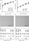
Fig. 1
Fig. 1
The effect of hFF on the general characteristics of placental MSCs. a The effect of three different concentrations (5, 10, and 20 %) of hFF on the proliferation rate of fPMSC-X. 105 fPMSC-X at P3 were plated on a 25 cm2 culture dish at the beginning of the experiment and trypan-blue negative cells were counted when the cells reached approximately 90 % confluency, which occurred at about day 3. Triplicate sets of experiments were repeated twice for each concentration of hFF. Results were expressed as mean ± standard error of mean. b The effect of hFF and FBS on the proliferation of placental MSCs, as revealed by the growth curves of fPMSC-X and fPMSC, respectively. Experimental sets and counts were performed as described above except for 48 days and results were expressed as mean ± standard error of mean. c General morphology of fPMSC-X and d fPMSC cultivated for approximately 60 days in vitro as shown under phase contrast (DIC) microscopy. Scale bars indicate 10 μm. e Karyotype analysis of fPMSC-X and f fPMSC grown for 60 days in vitro confirmed the normal chromosomal compositions and G-banding patterns
Fig. 2
Fig. 2
The effect of hFF on the gene expressions of placental MSCs. Expressions of Oct 4 (a, b), Nanog (c, d), and c-kit (e, f) were examined in fPMSC-X (a, c, e) and fPMSC (b, d, f) by immunocytochemistry and counterstaining with Hoechst 33342. Approximately 45.2 ± 2.3 % (a), 89.6 ± 1.6 % (c), 96.1 ± 1.0 % % (e) of fPMSC-X expressed Oct4, Nanog, and c-Kit, respectively, and 37.1 ± 2.0 % (b), 82.5 ± 1.3 % (d), 98.3 ± 0.5 % (f) of fPMSC expressed Oct4, Nanog, and c-Kit, respectively (Scale bar 30 μm) (g) RT-PCR analysis confirmed the expression of the pluripotency-coupled genes, Oct4, Nanog, Tbn, Activin, Klf4, and Rex1, which were well preserved in both culture conditions
Fig. 3
Fig. 3
The effect of hFF on the surface marker expressions of placental MSCs. FACS analysis revealed that fPMSC-X maintained in vitro for at least 60 days were positive for CD13, CD44, CD90, CD105 and HLA-ABC expression, but negative for CD14, CD34, CD45, CD127, and HLA-DR (a). The surface marker expression pattern was indistinguishable from those of fPMSC (b). The X axis of each diagram shows the relative fluorescence intensity of PE or FITC and the Y axis of each diagram shows the relative cell number
Fig. 4
Fig. 4
The effect of hFF on the differentiation potency of placental MSCs. Differentiation of fPMSC-X and fPMSC into endodermal and ectodermal cells. Approximately 30–50 % of undifferentiated fPMSC-X (a) and fPMSC (b) maintained for at least 48 days in vitro expressed nestin, and the percentage of the expression increased dramatically to 96.6 ± 2.4 and 94.9 ± 3.1 % in both fPMSC-X (c) and fPMSC (d), respectively, suggesting the ectodermal differentiation potency was well maintained in both of the culture conditions. The ectodermal differentiation tendency was further confirmed by RT-PCR (e). There was no spontaneous induction of insulin in either fPMSC-X (f) or in fPMSC (g) when cultivated for 48 days in their respective culture conditions. However, when the fPMSC-X (h) and fPMSC (i) were subjected to endodermal differentiation for 7–14 days, both types of cells spontaneously aggregated and expressed insulin as confirmed by immunohistochemistry. The endodermal differentiation was further confirmed by RT-PCR (j). Undiff denotes for fPMSC-X or fPMSC cultivated for at least 48 days in vitro and Diff denotes for the differentiated fPMSC-X or fPMSC. Scale bars indicate 30 μm

References

    1. Antoniades HN, Scher CD, Stiles CD. Purification of human platelet-derived growth factor. Proc Natl Acad Sci USA. 1979;76:1809–1813. doi: 10.1073/pnas.76.4.1809. - DOI - PMC - PubMed
    1. Attaran M, Pasqualotto E, Falcone T, Goldberg JM, Miller KF, Agarwal A, Sharma Rk. The effect of follicular fluid reactive oxygen species on the outcome of in vitro fertilization. Int J Fertil Womens Med. 2000;45:314–320. - PubMed
    1. Barlow S, Brook G, Chatterjee K, Price G, Pelekanos R, Rossetti T, Doody M, Venter D, Pain S, Gilshenan K, Atkinson K. Comparison of human placenta- and bonemarrow-derived multipotent mesenchymal stem cell. Stem Cells Dev. 2008;17:1095–1108. doi: 10.1089/scd.2007.0154. - DOI - PubMed
    1. Barnes D, Sato G. Methods for growth of cultured cells in serum-free medium. Anal Biochem. 1980;102:255–270. doi: 10.1016/0003-2697(80)90151-7. - DOI - PubMed
    1. Battula VL, Bareiss PM, Treml S, Conrad S, Albert I, Hojak S, Abele H, Schewe B, Just L, Skutella T, Bühring HJ. Human placenta and bone marrow derived MSC cultured in serum-free, b-FGF-containing medium express cell surface frizzled-9 and SSEA-4 and give rise to multilineage differentiation. Differentiation. 2007;75:279–291. doi: 10.1111/j.1432-0436.2006.00139.x. - DOI - PubMed

LinkOut - more resources