. 2014 Oct;10(10):4323-31.
doi: 10.1016/j.actbio.2014.06.034. Epub 2014 Jul 1.

Engineering alginate as bioink for bioprinting

Affiliations

Engineering alginate as bioink for bioprinting

Jia Jia et al. Acta Biomater. 2014 Oct.

Abstract

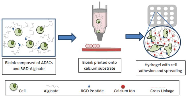
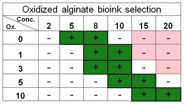
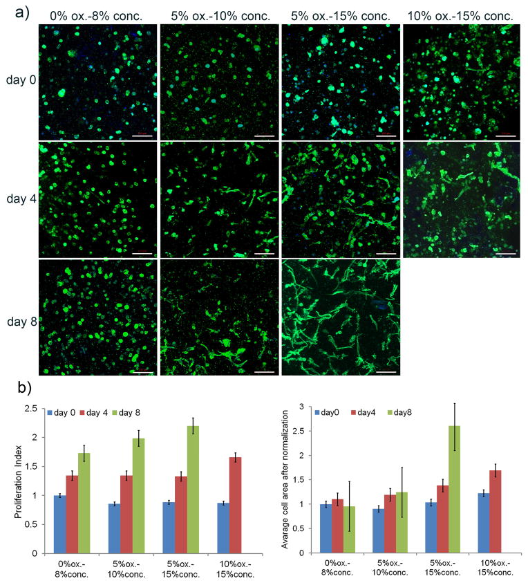
Recent advances in three-dimensional (3-D) printing offer an excellent opportunity to address critical challenges faced by current tissue engineering approaches. Alginate hydrogels have been used extensively as bioinks for 3-D bioprinting. However, most previous research has focused on native alginates with limited degradation. The application of oxidized alginates with controlled degradation in bioprinting has not been explored. Here, a collection of 30 different alginate hydrogels with varied oxidation percentages and concentrations was prepared to develop a bioink platform that can be applied to a multitude of tissue engineering applications. The authors systematically investigated the effects of two key material properties (i.e. viscosity and density) of alginate solutions on their printabilities to identify a suitable range of material properties of alginates to be applied to bioprinting. Further, four alginate solutions with varied biodegradability were printed with human adipose-derived stem cells (hADSCs) into lattice-structured, cell-laden hydrogels with high accuracy. Notably, these alginate-based bioinks were shown to be capable of modulating proliferation and spreading of hADSCs without affecting the structure integrity of the lattice structures (except the highly degradable one) after 8days in culture. This research lays a foundation for the development of alginate-based bioink for tissue-specific tissue engineering applications.

Keywords: Adipose-derived stem cells; Bioink; Bioprinting; Hydrogel scaffold; Oxidized alginate.


Figures

Figure 1
Figure 1
Schematic representation of biodegradable oxidized alginate as bioink for bioprinting. A bioink consisting of RGD-modified oxidized alginate hADSCs was printed in a define lattice structure on a gelatin substrate to crosslink the hydrogel. The constructs were then evaluated over an 8-day period for cellular behavior (i.e., cell proliferation and spreading).
Figure 2
Figure 2
Density-based analysis on printability of different alginate solutions. (a) Density (mean ± SD) measurements of each sample with successful cell suspension results (green). Red denotes alginate compositions that did not completely dissolve into solution after 2 days. The other materials (white) were unable to maintain a homogenous cell distribution. (b) Calcein-stained hADSCs in the 5% ox.-10% conc. (left) and 5% ox.-2% conc. (right) material with and without successful cell suspension, respectively (scale bar= 500 μm).
Figure 3
Figure 3
Viscosity-based analysis on printability of different alginate solutions. (a) Viscosity values of various alginate solutions with a range of concentrations and oxidation levels that passed the density requirement shown with a favorable area for higher resolution bioprinting with hADSCs (green). Red denotes alginate compositions that did not completely dissolved into solution after 2 days. The other materials were either too viscous to prepare for printing or failed the density test (white). (b) Using dots as the functional unit of liquid-dispensing strategies and a representation of resolution, a printed dot array (5×5) shows examples of low printing resolution (top), high printing resolution (low) (scale bar= 1 mm). (c) A plot of areas of dots versus viscosity shows a direct relationship between printability and viscosity of alginate samples. Guiding lines represent general flow of data.
Figure 4
Figure 4
Cell viability assay of density and viscosity criterion-filtered samples. (a) Samples of high viability (>90%) right after printing (green). (b) The fluorescent pictures of live-dead assay after printing: (i) high cell viability sample (e.g. 5% ox.-15% conc.) and (ii) low cell viability sample (e.g. 5% ox.-20% conc.) (scale bar= 100 μm). (c) Cell viability assay at day 8. Except the 5% ox.-20% conc. sample (0% viability), the remaining four samples showed high viabilities (>95%) after 8 days in culture (scale bar= 100 μm).
Figure 5
Figure 5
Summary table of the preferable range of alginate samples with high printability (green) based on the three established printability criteria (i.e., homogeneous cell suspension, high printing resolution, and high cell viability).
Figure 6
Figure 6
Lattice structures printed with bioprinting-compatible materials and their dimensional change in 8 days. (a) Initial design of lattice structure. (b) Pictures of printed lattice structures (5% ox.-10% conc. sample) at day 0, day 4, day 8 shows printed structures highly matched the initial design with apparent dimensional changes after 8 days in culture (scale bar= 2 mm). (c) Normalized comparison between the initial design (12.6 mm × 12.6 mm) and the X and Y dimensions of the lattice structures (0% ox.-8% conc., 5% ox.-10% conc., 5% ox.-15% conc.) after 8 days in culture. All values are mean ± SD. Asterisk denotes significant difference between day 0 and day 8.
Figure 7
Figure 7
hADSC behavior in the lattice structures. (a) Cell spreading with fluorescent staining (phalloidin) for actin at day 0, 4, and 8 (scale bar= 100 μm). (b) hADSC proliferation index and spreading assays based on fluorescent staining (phalloidin and DAPI stain) at day 8. Proliferation index was calculated as the cell number of each day divided by the original cell number on day 0 for the 0% ox.-8% conc. alginate sample for relative comparison. Cell area was calculated as total cell area divided by the number of cells normalized over the values at day 0 for the 0% ox.-8% conc. alginate sample. All values are mean ± SD.
Figure 8
Figure 8
A computer-rendered 3D picture of a portion of the printed lattice structure made by the best supporting hADSC material, 5% ox.-15% conc. oxidized alginate, showing multiple layers of spreading cells within the hydrogel.

References

    1. Visconti RP, Kasyanov V, Gentile C, Zhang J, Markwald RR, Mironov V. Towards organ printing: engineering an intra-organ branched vascular tree. Expert Opin Biol Ther. 2010;10:409–420. - PMC - PubMed
    1. Fedorovich NE, Alblas J, Hennink WE, Oner FC, Dhert WJA. Organ printing: the future of bone regeneration? Trends Biotechnol. 2011;29:601–606. - PubMed
    1. Mironov V, Kasyanov V, Markwald RR. Organ printing: from bioprinter to organ biofabrication line. Curr Opin Biotechnol. 2011;22:667–673. - PubMed
    1. Norotte C, Marga FS, Niklason LE, Forgacs G. Scaffold-free vascular tissue engineering using bioprinting. Biomaterials. 2009;30:5910–5917. - PMC - PubMed
    1. Seliktar D, Dikovsky D, Napadensky E. Bioprinting and tissue engineering: recent advances and future perspectives. Isr J Chem. 2013;53:1–10.

Publication types