Review
. 2014 Oct:30:60-7.
doi: 10.1016/j.ceb.2014.06.007. Epub 2014 Jul 5.

Self-organization of protrusions and polarity during eukaryotic chemotaxis

Affiliations
Review

Self-organization of protrusions and polarity during eukaryotic chemotaxis

Brian R Graziano et al. Curr Opin Cell Biol. 2014 Oct.

Abstract

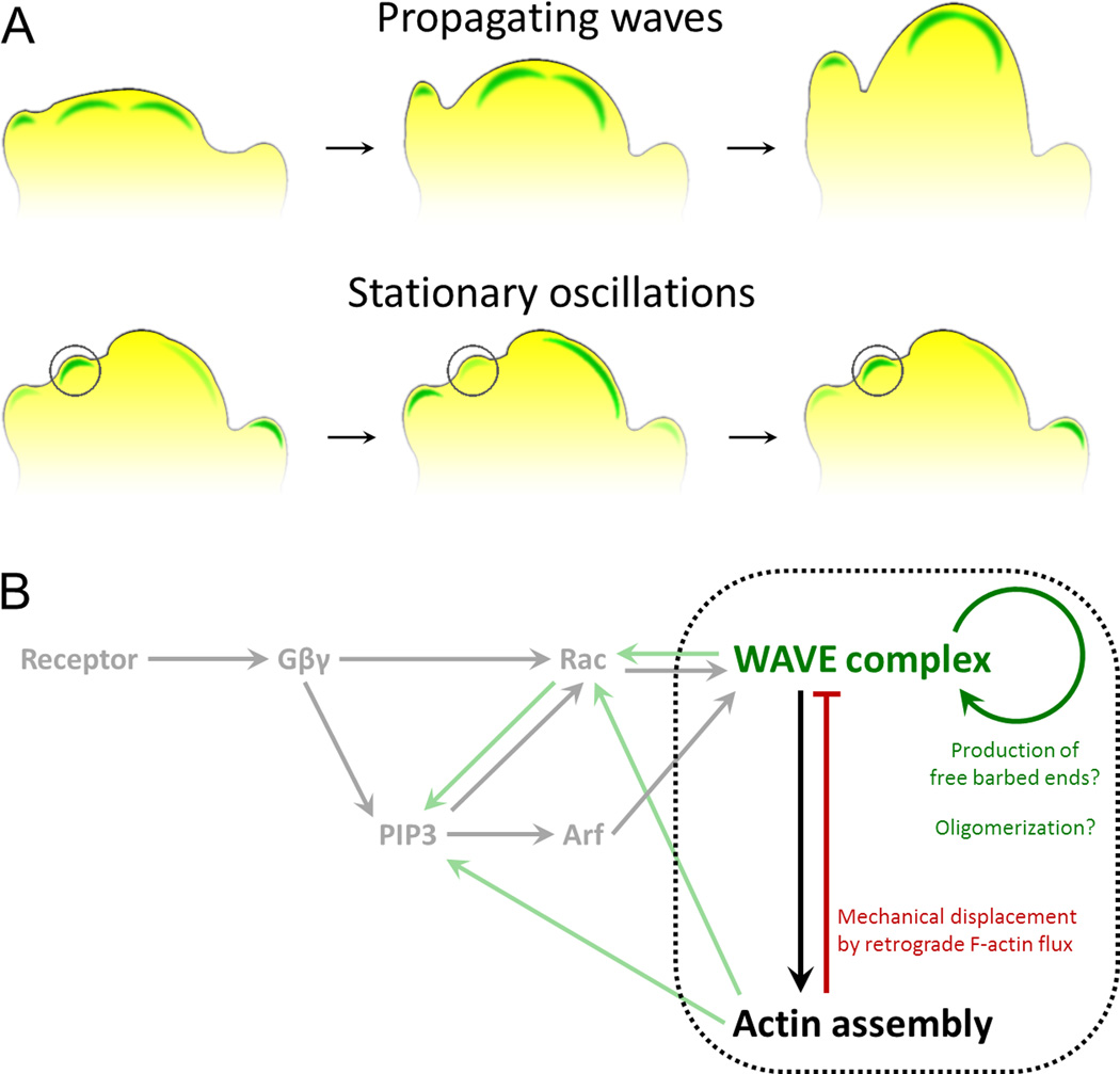
Many eukaryotic cells regulate their polarity and motility in response to external chemical cues. While we know many of the linear connections that link receptors with downstream actin polymerization events, we have a much murkier understanding of the higher order positive and negative feedback loops that organize these processes in space and time. Importantly, physical forces and actin polymerization events do not simply act downstream of chemotactic inputs but are rather involved in a web of reciprocal interactions with signaling components to generate self-organizing pseudopods and cell polarity. Here we focus on recent progress and open questions in the field, including the basic unit of actin organization, how cells regulate the number and speed of protrusions, and 2D versus 3D migration.


Figures

Figure 1
Figure 1. Actin polymerization as an excitable system
A) Actin polymerization is organized as multiple wavelike (top) and oscillatory nucleation events (bottom, single oscillator denoted by circle) in motile cells, as visualized by the dynamics of the Wave Regulatory Complex (WRC) [–11]. Waves that reach the cell boundary organize cellular protrusion. B) The behavior of these waves and oscillations are consistent with an excitable system, which typically has the topology of rapid positive feedback and delayed inhibition (components bound by black dotted line). Key components of the chemotactic signaling network and the flow of information to factors specifying actin assembly are depicted in gray. Candidates for the molecular basis of the positive and negative feedback loops (green and red arrows, respectively) are briefly summarized.
Figure 2
Figure 2. Actin assembly reciprocally interacts with mechanical forces to regulate protrusion number and speed
A) In the presence of uniform chemoattractant, cells form a single leading edge through competition between nascent protrusions. This competition occurs through long-range inhibition that is communicated by changes in membrane tension. Actin polymerization at each protrusion exerts force on the plasma membrane (grey arrows), rapidly increasing membrane tension (black double-arrows) throughout the cell. Increased membrane tension globally suppresses the formation of new sites of polarized actin assembly through inhibition of upstream signals such as Rac activation and WRC recruitment; however, assembly still occurs at the initial site since positive feedback components are already present at sufficiently high levels to overcome this inhibition. This system ensures that a single dominant leading edge emerges [66], but it is not yet clear how changes in tension are translated to changes in signaling. B) Dendritic cells in a confined environment maintain a steady rate of forward protrusion, independent of surface adhesiveness. To compensate for reduced traction on non-adhesive surfaces and the subsequent increase in the rate of retrograde flow of the actin network (green arrows), cells increase the rate of actin polymerization directed toward the leading edge (grey arrows) to produce the same protrusion velocity of the plasma membrane (black arrows). Even a single cell that finds itself overlapping surfaces with differing levels of adhesiveness is able to maintain distinct subcellular domains with different rates of actin assembly to produce the same net force across the leading edge and maintain persistence despite the adhesive differences. These data suggest that the speed of cell protrusion is not set by the amount of actin assembly but rather depends on how fast the plasma membrane is released into the protrusions [52]. How this is regulated is unknown.

References

    1. De Bruyn PP. The amoeboid movement of the mammalian leukocyte in tissue culture. Anat Rec. 1946;95:177–191. - PubMed
    1. Hartman RS, Lau K, Chou W, Coates TD. The fundamental motor of the human neutrophil is not random: evidence for local non-Markov movement in neutrophils. Biophys J. 1994;67:2535–2545. - PMC - PubMed
    1. Killich T, Plath PJ, Wei X, Bultmann H, Rensing L, Vicker MG. The locomotion, shape and pseudopodial dynamics of unstimulated Dictyostelium cells are not random. J Cell Sci. 1993;106(Pt 4):1005–1013. Through careful quantitative measure of cell shape and pseudopod dynamics, this paper provides evidence that the actin cytoskeleton is an oscillatory system more than a decade before fluorescence imaging enabled direct visualization of oscillatory and wavelike actin dynamics.

    1. Vicker MG. Reaction-diffusion waves of actin filament polymerization/depolymerization in Dictyostelium pseudopodium extension and cell locomotion. Biophys Chem. 2000;84:87–98. - PubMed
    1. Vicker MG. Eukaryotic cell locomotion depends on the propagation of self-organized reaction-diffusion waves and oscillations of actin filament assembly. Exp Cell Res. 2002;275:54–66. - PubMed

Publication types

LinkOut - more resources