Unusual case of recurrent thigh lump in a girl: a case report
- PMID: 24998416
- PMCID: PMC4105883
- DOI: 10.1186/1752-1947-8-245
Unusual case of recurrent thigh lump in a girl: a case report
Abstract
Introduction: Lipofibromatosis is a rare fibro-fatty tumour with a predilection to involve distal extremities. It has only recently been described as a distinctive clinicopathologic entity, and subsequently only a few cases have been published in the literature. To address the clinicopathologic significance of this rare entity, we here describe a case of lipofibromatosis occurring on the left thigh of a Sri Lankan girl who developed a recurrence following excision.
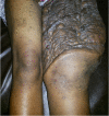
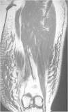
Case presentation: A 15-year-old previously healthy girl of Sri Lankan ethnicity presented with a painless progressively enlarging mass in her left thigh. Magnetic resonance imaging of her thigh lump, revealed a septated mass arising from subcutaneous tissue of anterolateral and medial aspects of her thigh. Histological assessment revealed evidence of lipofibromatosis, and the lesion was excised followed by split-skin grafting. She presented again with a local recurrence at the same site.
Conclusions: Adequate surgical excision leads to complete cure of this benign lesion, but recurrences are common following incomplete excision. Therefore awareness among clinicians of this rare entity is vital in offering the best possible care to the patients.
Figures


Similar articles
-
Lipofibromatosis: A Rare Diagnosis on Fine Needle Aspiration Cytology.Turk Patoloji Derg. 2020;1(1):268-274. doi: 10.5146/tjpath.2020.01479. Turk Patoloji Derg. 2020. PMID: 32149363 Free PMC article.
-
Hibernoma of the thigh: a lipoma-like variant rare tumour mimicking soft tissue sarcoma.BMJ Case Rep. 2012 Nov 30;2012:bcr2012007315. doi: 10.1136/bcr-2012-007315. BMJ Case Rep. 2012. PMID: 23203180 Free PMC article.
-
Lipofibromatosis of the knee in a 19-month-old child.J Pediatr Surg. 2012 May;47(5):1028-31. doi: 10.1016/j.jpedsurg.2012.02.009. J Pediatr Surg. 2012. PMID: 22595596
-
Lipofibromatosis: an institutional and literature review of an uncommon entity.Pediatr Dermatol. 2014 May-Jun;31(3):298-304. doi: 10.1111/pde.12335. Pediatr Dermatol. 2014. PMID: 24758203 Review.
-
[Desmoid fibroma of soft tissues of extra-abdominal sites: review of the literature apropos of a case located at the posterior face of the thigh].Ann Chir. 1989;43(4):289-94. Ann Chir. 1989. PMID: 2660722 Review. French.
References
-
- Fetsch JF, Miettinen M, Laskin WB, Michal M, Enzinger FM. A clinicopathologic study of 45 pediatric soft tissue tumors with an admixture of adipose tissue and fibroblastic elements, and a proposal for classification as lipofibromatosis. Am J Surg Pathol. 2000;24(11):1491–1500. doi: 10.1097/00000478-200011000-00004. - DOI - PubMed
Publication types
MeSH terms
LinkOut - more resources
Full Text Sources
Other Literature Sources

