Case Reports
. 2015;55(8):683-8.
doi: 10.2176/nmc.tn.2013-0249. Epub 2014 Jul 4.

Fluorescence Angiography with Temporary Occlusion to Confirm the Distal Artery: Technical Notes

Affiliations
Case Reports

Fluorescence Angiography with Temporary Occlusion to Confirm the Distal Artery: Technical Notes

Yasuo Murai et al. Neurol Med Chir (Tokyo). 2015.

Abstract

Confirming the patency of the proximal parent and distal artery is necessary in cerebral aneurysm surgery. To understand the relationship between the parent and distal arteries of the aneurysm, the blood vessels running through the subarachnoid space should be extensively dissected, which is time consuming. To examine the efficacy of a temporary clip with indocyanine green (ICG) technique, in which the parent artery is temporarily occluded using a temporary clip, an ICG videoangiography (ICGVAG) is performed to clarify the relationship between the distal artery and the proximal parent artery. Three patients with a distal aneurysm. This technique was used to confirm the connection of the parent and the distal artery in distal aneurysms. With regard to middle cerebral artery (MCA), the procedure is conducted as follows. First, the M2 within the Sylvian fissure is investigated to ensure the absence of atherosclerosis and perforators and that this vessel could undergo occlusion by temporary clipping. The subarachnoid space surrounding the distal artery of the lesion site suspected of an existent aneurysm is dissected. The image range of the ICGVAG is set sufficiently wide to accommodate the possibility that the distal artery is not the artery that was anticipated. Subsequently, after the temporary clip occlusion is completed, the ICGVAG is recorded. In the three distal aneurysms, the relationship between the aneurysm, the distal artery, and the parent artery was confirmed. This method was useful, suggesting that unnecessary dissection in the subarachnoid space might be reduced.


Conflict of interest statement

Conflicts of Interest Disclosure

The authors declare no conflicts of interest. All authors declare no personal interest in any of the materials or devices in this article.

Figures

Fig. 1.
Fig. 1.
Left: Initial preoperative three-dimensional computed tomography (CT) angiography demonstrating a distal posterior inferior cerebellar artery aneurysm on the cortical segment. The posterior inferior cerebellar artery and the anterior inferior cerebellar artery were branching from almost the identical segment of the vertebral artery. Right: The post-operative lateral view of the left vertebral digital subtraction angiography showing good patency of the distal posterior inferior cerebellar artery and no residual aneurysm is seen.
Fig. 2.
Fig. 2.
Left: The microscopic findings of the surgical field. The tonsillomedullary segment of the left posterior inferior cerebellar artery was temporary clipped. Middle: An ICGVAG image taken 16 s after the ICG infusion. The peripheral left posterior inferior cerebellar artery (white arrow) was enhanced from the collateral circulation. The proximal side of the posterior inferior cerebellar artery was not enhanced (arrowhead), because of the temporary clip. Right: An ICGVAG image taken 22 s after the ICG infusion just after the removal of the temporary clip. The enhancement of the peripheral left posterior inferior cerebellar artery (arrowhead) was diminished (white arrow) just after the removal (arrowhead) of the clip. ICGVAG: indocyanine green videoangiography.
Fig. 3.
Fig. 3.
The microscopic findings of the aneurysm after clipping. The white arrow indicates the proximal artery, and the white arrowhead indicates the distal artery.
Fig. 4.
Fig. 4.
Intraoperative ICGVAG images. Image A was captured 11 s after the ICG infusion before the removal of the temporary clip. The frontal peripheral middle cerebral artery (arrowhead) has been thinly enhanced. The temporal peripheral middle cerebral artery (arrow) has been thickly enhanced. Image B was captured just before the removal of temporary clip. The frontal peripheral middle cerebral artery (arrowhead) and the temporal peripheral middle cerebral artery (arrow) have been enhanced at the same level. Image C was captured just after the removal of the temporary clip. The enhancement of the frontal peripheral middle cerebral artery (arrowhead) was diminished just after the removal of the clip. ICGVAG: indocyanine green videoangiography.
Fig. 5.
Fig. 5.
Fluorescent intensity curve with the Carl Zeiss FLOW 800 system (Carl Zeiss Co., Tokyo). The pink curve indicates the frontal peripheral middle cerebral artery and the red curve indicates the frontal peripheral middle cerebral artery. The blue circle on the pink curve indicates a temporary change of intensity just after the removal of the temporary clip.
Fig. 6.
Fig. 6.
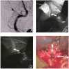
Upper left: Anterior posterior view of the initial digital subtraction angiography demonstrating a distal posterior inferior cerebellar artery aneurysm on the cortical segment. Upper right: An ICGVAG image taken 22 s after ICG infusion. The peripheral left posterior inferior cerebellar artery (white arrow) vessel has been thinly enhanced. Lower left: An ICGVAG image just after the removal of the temporary clip. The strength of the enhancement of the peripheral left PICA (white arrow) was changed. Lower right: The microscopic findings of the aneurysm before clipping. The white arrow indicates the proximal artery, and the white arrowhead indicates the aneurysm. ICGVAG: indocyanine green videoangiography, PICA: posterior inferior cerebellar artery.

Similar articles

Cited by

References

    1. Dashti R, Hernesniemi J, Niemelä M, Rinne J, Lehecka M, Shen H, Lehto H, Albayrak BS, Ronkainen A, Koivisto T, Jääskeläinen JE: Microneurosurgical management of distal middle cerebral artery aneurysms. Surg Neurol 67: 553– 563, 2007. - PubMed
    1. Horiuchi T, Tanaka Y, Takasawa H, Murata T, Yako T, Hongo K: Ruptured distal middle cerebral artery aneurysm. J Neurosurg 100: 384– 388, 2004. - PubMed
    1. Horiuchi T, Tanaka Y, Hongo K, Nitta J, Kusano Y, Kobayashi S: Characteristics of distal posteroinferior cerebellar artery aneurysms. Neurosurgery 53: 589– 595; discussion 595–596, 2003. - PubMed
    1. Joo SP, Kim TS, Choi JW, Lee JK, Kim YS, Moon KS, Kim JH, Kim SH: Characteristics and management of ruptured distal middle cerebral artery aneurysms. Acta Neurochir (Wien) 149: 661–667, 2007. - PubMed
    1. Lee SH, Bang JS: Distal middle cerebral artery M4 aneurysm surgery using navigation-CT angiography. J Korean Neurosurg Soc 42: 478– 480, 2007. - PMC - PubMed

Publication types