

Morphological and immunohistochemical study of ovarian and tubal dysplasia associated with tamoxifen
- PMID: 24998918
- PMCID: PMC4083318
- DOI: 10.4081/ejh.2014.2251
Morphological and immunohistochemical study of ovarian and tubal dysplasia associated with tamoxifen
Abstract
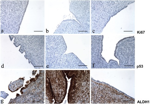
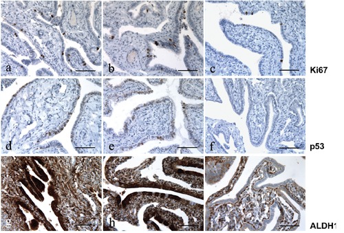
Ovarian epithelial dysplasia was initially described in material from prophylactic oophorectomies for BReast CAncer gene (BRCA) mutation. Similar histopathological abnormalities have been revealed after ovulation stimulation. Given that tamoxifen (TAM) has a clomid-like effect and is sometimes used to induce ovulation, we studied the morphological features and immunohistochemical expression patterns of neoplasia-associated markers in adnexectomies previously exposed to TAM for breast cancer. We blindly reviewed 173 histopathological slides of adnexectomies according to three groups - oophorectomie sassociated with TAM exposure (n=42), oophorectomies associated with clomiphene exposure (n=15) and a spontaneously fertile non cancerous control group (n=116). Morphological features (with an ovarian and tubal dysplasia scoring system) and immunohistochemical expression patterns of Ki-67, p53 and Aldehyde dehydrogenase 1 (ALDH1 is an enzyme significantly associated with earlystage ovarian cancer) were evaluated and correlated. Mean tubal dysplasia score was significantly higher in the TAM group and clomiphene group than in controls (respectively 7.8 vs 3.5, P<0.007 and 6.8 vs 3.5, P=0.008). There is no statistical difference for the ovarian score in TAM group in comparison with the control group whereas we found a significant score for clomiphen group (6.5, P=0.009). Increased ALDH1 expression was observed in the two exposed group whereas expression patterns of Ki67 and p53 were moderate. Interestingly, ALDH1 expression was low in non-dysplastic epithelium, high in dysplasia, and constantly low in the two carcinoma. Furthermore, we confirm our previous results showing that ALDH1 may be a useful tissue biomarker in the subtle histopathological diagnosis of tubo-ovarian dysplasia.
Conflict of interest statement
Conflict of interests: the authors declare no conflict of interests.
Figures
Similar articles
-
Morphological and immunohistochemical pattern of tubo-ovarian dysplasia and serous tubal intraepithelial carcinoma.Eur J Obstet Gynecol Reprod Biol. 2014 Dec;183:89-95. doi: 10.1016/j.ejogrb.2014.10.003. Epub 2014 Oct 14. Eur J Obstet Gynecol Reprod Biol. 2014. PMID: 25461359
-
Early telomere shortening and genomic instability in tubo-ovarian preneoplastic lesions.Clin Cancer Res. 2013 Jun 1;19(11):2873-82. doi: 10.1158/1078-0432.CCR-12-3947. Epub 2013 Apr 15. Clin Cancer Res. 2013. PMID: 23589176
-
[Morphologic changes of fallopian tubal epithelium in ovarian serous tumors].Zhonghua Bing Li Xue Za Zhi. 2012 Jul;41(7):433-7. doi: 10.3760/cma.j.issn.0529-5807.2012.07.001. Zhonghua Bing Li Xue Za Zhi. 2012. PMID: 22932451 Chinese.
-
Ovarian endometrioid adenocarcinoma arising from an endometriotic cyst in a postmenopausal woman under tamoxifen therapy for breast cancer: a case report.Gynecol Oncol. 2002 Nov;87(2):231-4. doi: 10.1006/gyno.2002.6824. Gynecol Oncol. 2002. PMID: 12477460 Review.
-
Uterine neoplasms in patients treated with tamoxifen.J Cell Biochem Suppl. 1995;23:179-83. doi: 10.1002/jcb.240590924. J Cell Biochem Suppl. 1995. PMID: 8747394 Review.
Cited by
-
Expression of Stem Cell Markers in Preinvasive Tubal Lesions of Ovarian Carcinoma.Biomed Res Int. 2015;2015:808531. doi: 10.1155/2015/808531. Epub 2015 Oct 4. Biomed Res Int. 2015. PMID: 26504831 Free PMC article.
-
The Importance of Steroid Uptake and Intracrine Action in Endometrial and Ovarian Cancers.Front Pharmacol. 2017 Jun 19;8:346. doi: 10.3389/fphar.2017.00346. eCollection 2017. Front Pharmacol. 2017. PMID: 28674494 Free PMC article. Review.
-
Is there still room for novelty, in histochemical papers?Eur J Histochem. 2016 Dec 16;60(4):2758. doi: 10.4081/ejh.2016.2758. Eur J Histochem. 2016. PMID: 28076939 Free PMC article. Review.
-
Genome-wide transcriptional regulation of estrogen receptor targets in fallopian tube cells and the role of selective estrogen receptor modulators.J Ovarian Res. 2016 Feb 15;9:5. doi: 10.1186/s13048-016-0213-3. J Ovarian Res. 2016. PMID: 26879975 Free PMC article.
-
Histochemistry in biology and medicine: a message from the citing journals.Eur J Histochem. 2015 Dec 23;59(4):2610. doi: 10.4081/ejh.2015.2610. Eur J Histochem. 2015. PMID: 26708189 Free PMC article. Review.
References
-
- Brown J, Farquhar C, Beck J, Boothroyd C, Hugues E. Clomiphene and anti-oestrogens for ovulation induction in PCOS. Cochrane Database Syst Rev 2009;4: CD002249. - PubMed
-
- Badawy A, Gibreal A. Clomiphene citrate versus tamoxifen for ovulation induction in women with PCOS : a prospective randomized trial. Eur J Obstet Gynecol Reprod Biol 2011;159: 151-4 - PubMed
-
- Salazar H, Godwin AK, Daly MB, Laub PB, Hogan M, Rosenblum N, et al. Microscopic benign and invasive malignant neoplasms and a cancer-prone phenotype in prophylactic oophorectomies. J Natl Cancer Inst 1996;88: 1810-20 - PubMed
-
- Werness BA, Afify AM, Bielat KL, Eltabbakh GH, Piver MS, Paterson JM. Altered surface and cyst epithelium of ovaries removed prophylactically from women with a family history of ovarian cancer. Hum Pathol 1999;30: 151-7 - PubMed
-
- Stratton JF, Buckey CH, Lowe D, Ponder BAJ. Comparison of prophylactic oophorectomy specimens from carriers and non carriers of a BRCA1 or BRCA2 gene mutation. J Natl Cancer Inst 1999;91: 626-8 - PubMed
Publication types
MeSH terms
Substances
LinkOut - more resources
Full Text Sources
Other Literature Sources
Medical
Research Materials
Miscellaneous
