

Expressions of local renin-angiotensin system components in chondrocytes
- PMID: 24998927
- PMCID: PMC4083327
- DOI: 10.4081/ejh.2014.2387
Expressions of local renin-angiotensin system components in chondrocytes
Abstract
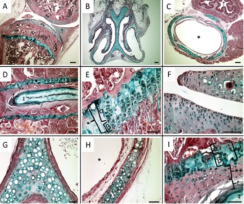
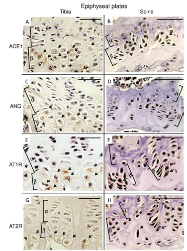
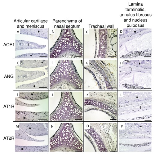
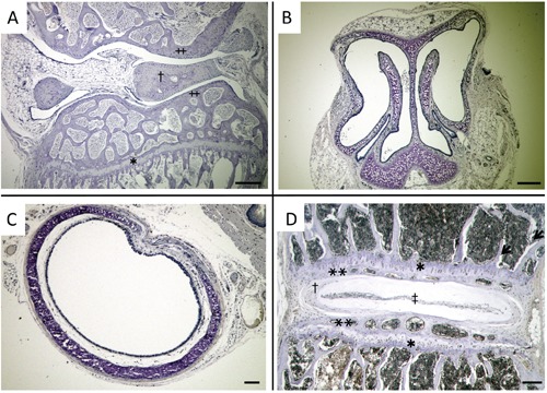
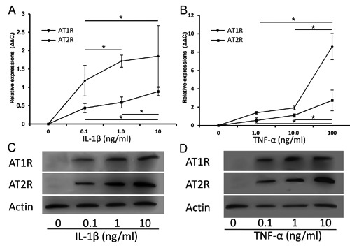
In 2013, we reported that local renin-angiotensin system (local RAS) components express during the hypertrophic differentiation of chondrocytes and can modulate it, using ATDC5 cell line that involves differentiation from mesenchymal stem cells to calcified hypertrophic chondrocytes. However, the expressions of local RAS components in normal chondrocytes have not been revealed yet. The purpose of this study is to examine the expression of the local RAS components in chondrocytes in vivo and the conditions allowing the expression. We stained five major regions of 8-week-old C57BL/6 adult mice in which chondrocytes exist, including epiphyseal plates and hyaline cartilages, with antibodies to local RAS components. We also examined the expression of local RAS components in the cultured bovine's articular cartilage chondrocytes using quantitative reverse transcription polymerase chain reaction and western blot analysis. In result, hypertrophic chondrocytes of epiphyseal plates included in the tibia and the lamina terminals expressed local RAS components. However, hyaline chondrocytes, including the knee articular cartilages, the parenchyma of nasal septums and of the tracheal walls, did not express local RAS components. Cultured bovine's articular cartilage chondrocytes also did not express local RAS components. However, inducing hypertrophy by administering interleukin-1β or tumor necrosis factor-α, the cultured articular chondrocytes also expressed angiotensin II type 1 receptor and angiotensin II type 2 receptor. In conclusion, local RAS components express particularly in chondrocytes which occur hypertrophy and do not in hyaline chondrocytes. The results are in accord with our previous in vitro study. We think this novel knowledge is important to investigate cartilage hypertrophy and diseases induced by hypertrophic changes like osteoarthritis.
Figures
Similar articles
-
Aliskiren has chondroprotective efficacy in a rat model of osteoarthritis through suppression of the local renin-angiotensin system.Mol Med Rep. 2017 Oct;16(4):3965-3973. doi: 10.3892/mmr.2017.7110. Epub 2017 Jul 28. Mol Med Rep. 2017. PMID: 28765966 Free PMC article.
-
Continuous infusion of angiotensin II modulates hypertrophic differentiation and apoptosis of chondrocytes in cartilage formation in a fracture model mouse.Hypertens Res. 2015 Jun;38(6):382-93. doi: 10.1038/hr.2015.18. Epub 2015 Feb 19. Hypertens Res. 2015. PMID: 25693858
-
Connective tissue growth factor mRNA expression pattern in cartilages is associated with their type I collagen expression.Bone. 2003 Dec;33(6):911-8. doi: 10.1016/j.bone.2003.07.010. Bone. 2003. PMID: 14678850
-
Regulation of energy metabolism in the growth plate and osteoarthritic chondrocytes.Rheumatol Int. 2018 Nov;38(11):1963-1974. doi: 10.1007/s00296-018-4103-4. Epub 2018 Jul 17. Rheumatol Int. 2018. PMID: 30019225 Review.
-
Renin-angiotensin system in osteoarthritis: A new potential therapy.Int Immunopharmacol. 2019 Oct;75:105796. doi: 10.1016/j.intimp.2019.105796. Epub 2019 Aug 10. Int Immunopharmacol. 2019. PMID: 31408841 Review.
Cited by
-
The role of oxidation of low-density lipids in pathogenesis of osteoarthritis: A narrative review.J Int Med Res. 2020 Jun;48(6):300060520931609. doi: 10.1177/0300060520931609. J Int Med Res. 2020. PMID: 32552129 Free PMC article. Review.
-
Current understanding of the link between angiotensin-converting enzyme and pain perception.Drug Discov Today. 2024 Sep;29(9):104089. doi: 10.1016/j.drudis.2024.104089. Epub 2024 Jul 6. Drug Discov Today. 2024. PMID: 38977123 Free PMC article. Review.
-
Increased H3K27ac level of ACE mediates the intergenerational effect of low peak bone mass induced by prenatal dexamethasone exposure in male offspring rats.Cell Death Dis. 2018 May 29;9(6):638. doi: 10.1038/s41419-018-0701-z. Cell Death Dis. 2018. PMID: 29844424 Free PMC article.
-
[Expressions of Renin, angiotensin converting enzyme, angiotensin receptor 1, and angiotensin receptor 2 in synovial tissue of osteoarthritis at different stages].Zhongguo Xiu Fu Chong Jian Wai Ke Za Zhi. 2020 Mar 15;34(3):362-366. doi: 10.7507/1002-1892.201904065. Zhongguo Xiu Fu Chong Jian Wai Ke Za Zhi. 2020. PMID: 32174084 Free PMC article. Chinese.
-
Co-Expression and Co-Localization of Cartilage Glycoproteins CHI3L1 and Lubricin in Osteoarthritic Cartilage: Morphological, Immunohistochemical and Gene Expression Profiles.Int J Mol Sci. 2016 Mar 11;17(3):359. doi: 10.3390/ijms17030359. Int J Mol Sci. 2016. PMID: 26978347 Free PMC article.
References
-
- Tigerstedt R, Bergman PG. Niere und Kreislauf. Skand Arch Physiol. 1898;8:223-71
-
- Marks LS, Maxwell MH. Tigerstedt and the discovery of renin. An historical note. Hypertension 1979;1:384-8 - PubMed
-
- Paul M, Poyan Mehr A, Kreutz R. Physiology of local renin-angiotensin systems. Physiol Rev. 2006;86:747-803 - PubMed
-
- Paul M, Bader M, Steckelings UM, Voigtländer T, Ganten D. The reninangiotensin system in the brain. Localization and functional significance. Arzneimittelforschung. 1993;43:207-13 - PubMed
-
- Zhao W, Leung PY, Chew SB, Chan HC, Wong PY. Localization and distribution of angiotensin II in the rat epididymis J. Endocrinol. 1996;149:217-22 - PubMed
Publication types
MeSH terms
Substances
LinkOut - more resources
Full Text Sources
Other Literature Sources
