. 2014 Jul 7:4:5593.
doi: 10.1038/srep05593.

One-step shell polymerization of inorganic nanoparticles and their applications in SERS/nonlinear optical imaging, drug delivery, and catalysis

Affiliations

One-step shell polymerization of inorganic nanoparticles and their applications in SERS/nonlinear optical imaging, drug delivery, and catalysis

Tzu-Ming Liu et al. Sci Rep. .

Abstract

Surface functionalized nanoparticles have found their applications in several fields including biophotonics, nanobiomedicine, biosensing, drug delivery, and catalysis. Quite often, the nanoparticle surfaces must be post-coated with organic or inorganic layers during the synthesis before use. This work reports a generally one-pot synthesis method for the preparation of various inorganic-organic core-shell nanostructures (Au@polymer, Ag@polymer, Cu@polymer, Fe3O4@polymer, and TiO2@polymer), which led to new optical, magnetic, and catalytic applications. This green synthesis involved reacting inorganic precursors and poly(styrene-alt-maleic acid). The polystyrene blocks separated from the external aqueous environment acting as a hydrophobic depot for aromatic drugs and thus illustrated the integration of functional nanoobjects for drug delivery. Among these nanocomposites, the Au@polymer nanoparticles with good biocompatibility exhibited shell-dependent signal enhancement in the surface plasmon resonance shift, nonlinear fluorescence, and surface-enhanced Raman scattering properties. These unique optical properties were used for dual-modality imaging on the delivery of the aromatic photosensitizer for photodynamic therapy to HeLa cells.


Figures

Figure 1
Figure 1
Schemes illustrated the polymer-assisted reduction of the HAuCl4 precursor to prepare Au@polymer NPs a), which exhibited polymer shell-dependent SERS and nonlinear optical enhancements for the observation of microscopic drug delivery in system b). c) The synthesis strategy using the different metal salt precursors to prepare Ag@polymer, Cu@polymer, TiO2@polymer, and Fe3O4@polymer NPs.
Figure 2
Figure 2
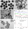
TEM images of the Au NPs prepared by the reaction of 0.25-mL a), 0.5-mL b), and 1-mL c) HAuCl4 (5 mM) with 10 mL of PSMA polymer (0.78 mg/mL) at 200°C for 1 h. d) UV-visible spectra for these corresponding Au NPs.
Figure 3
Figure 3
Time-dependent examinations of the a-c) TEM images from 1 h to 6 h to 13 h and d) UV-visible absorption spectra for the as-prepared Au@polymer NPs (~22 nm in core size). e) Au core size/polymer shell thickness plots f) Synchrotron X-ray powder diffraction pattern of the 13 h sample of the Au@polymer NPs. g) Raman spectra of the 1, 6, and 13 h samples of the Au@polymer NPs, where the * symbol shows the styrene portion of PSMA and the D/G bands refer to the sp3/sp2 hybridized carbon atoms of the graphite structure.
Figure 4
Figure 4. a) Nonlinear optical spectra of the as-prepared Au@polymer NPs at different reaction times and b) Raman shift detection of MB-tagged Au@polymer NPs (prepared over 1–13 h) at an excitation of 632.8 nm.
Figure 5
Figure 5. UV-visible and fluorescence spectra of different aromatic drugs encapsulated in the Au@polymer NPs (6 h-sample).
Figure 6
Figure 6
a) MTT assay of HeLa cells with free MB molecules and MB-loaded Au@polymer NPs at 24 h. b) Bright-field image of the HeLa cells with MB-loaded Au@polymer NPs and two SERS mapping images (based on the 1623 cm−1 vibration of MB) collected for the rectangle shown in the bright field of a HeLa cell. N indicates the nucleus. c) Fluorescence images of DCFH-DA staining in HeLa cells incubated with MB-loaded Au@polymer NPs and free MB for 4 h. The sample-treated cells were exposed to LED light at 660 nm (30 mW/cm2) for 4 min before imaging detection. d) Fluorescence intensity of MB-loaded Au@polymer NPs and free MB in HeLa cells.
Figure 7
Figure 7
TEM images of a) Ag@polymer (with 12 μL HCl and 100 μL N2H4), b) Cu@polymer (with 18 μL HCl and 100 μL N2H4), c) Fe3O4@polymer (with 100 μL N2H4), and d) TiO2@polymer (with 18 μL HCl and 100 μL N2H4) NPs. e) XRD measurements for these inorganic-polymer nanocomposites going from a) the bottom to d) the top. The peaks for a), b), c), and d) were assigned based on the standard patterns of Ag (JCPDS 04-0783), Cu (JCPDS 85-1326), Fe3O4 (JCPDS 19-629), and TiO2 (JCPDS 89-4921), respectively. f) UV-visible spectra of Ag@polymer and Cu@polymer NPs.

Similar articles

Cited by

References

    1. Huschka R., Neumann O., Barhoumi A. & Halas N. J. Visualizing light-triggered release of molecules inside living cells. Nano Lett. 10, 4117–4122 (2010). - PMC - PubMed
    1. Kang H. et al. Near-infrared light-responsive core–shell nanogels for targeted drug delivery. ACS Nano 5, 5094–5099 (2011). - PMC - PubMed
    1. Doane T. L. & Burda C. The unique role of nanoparticles in nanomedicine: imaging, drug delivery and therapy. Chem. Soc. Rev. 41, 2885–2911 (2012). - PubMed
    1. Jin Y. Engineering plasmonic gold nanostructures and metamaterials for biosensing and nanomedicine. Adv. Mater. 24, 5153–5165 (2012). - PubMed
    1. Yong K. T. Quantum dots for biophotonics. Theranostics 2, 629–630 (2012). - PMC - PubMed

Publication types

MeSH terms