TGF-β1, IL-6, and TNF-α in bronchoalveolar lavage fluid: useful markers for lung cancer?
- PMID: 24999009
- PMCID: PMC4083430
- DOI: 10.1038/srep05595
TGF-β1, IL-6, and TNF-α in bronchoalveolar lavage fluid: useful markers for lung cancer?
Abstract
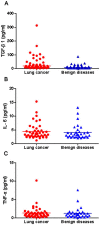
Changes of cytokines in bronchoalveolar lavage fluid (BALF) reflect immunologic reactions of the lung in pulmonary malignancies. Detection of biomarkers in BALF might serve as an important method for differential diagnosis of lung cancer. A total of 78 patients admitted into hospital with suspected lung cancer were included in our study. BALF samples were obtained from all patients, and were analyzed for TGF-β1, IL-6, and TNF-α using commercially available sandwich ELISA kits. The levels of TGF-β1 in BALF were significantly higher in patients with lung cancer compared with patients with benign diseases (P = 0.003). However, no significant difference of IL-6 (P = 0.61) or TNF-α (P = 0.72) in BALF was observed between malignant and nonmalignant groups. With a cut-off value of 10.85 pg/ml, TGF-β1 showed a sensitivity of 62.2%, and a specificity of 60.6%, in predicting the malignant nature of pulmonary disease. Our data suggest that TGF-β1 in BALF might be a valuable biomarker for lung cancer. However, measurement of IL-6 or TNF-α in BALF has poor diagnostic value in lung cancer.
Figures


References
-
- Sozzi G. et al. Detection of microsatellite alterations in plasma DNA of lung cancer patients: a prospect for early diagnosis. Clinical. Cancer. Research. 5, 2689–92 (1999). - PubMed
-
- Wingo P. A. et al. Annual report to the nation on the status of cancer, 1973–1996, with a special section on lung cancer and tobacco smoking. J. Natl. Cancer. Inst. 9, 675–90 (1999). - PubMed
-
- Ellis J. R. & Gleeson F. V. Lung cancer screening. J. Br. J. Radiol. 74, 478–85 (2001). - PubMed
-
- Marcus P. M. Lung cancer screening: an update. J. Clin. Oncol. 19, 83S–6S (2001). - PubMed
-
- Wang P., Piao Y., Zhang X., Li W. & Hao X. The concentration of CYFRA 21-1, NSE and CEA in cerebro-spinal fluid can be useful indicators for diagnosis of meningeal carcinomatosis of lung cancer. Cancer. Biomark. 13, 123–30 (2013). - PubMed
Publication types
MeSH terms
Substances
LinkOut - more resources
Full Text Sources
Other Literature Sources
Medical
Miscellaneous

