

Prenatal cocaine effects on brain structure in early infancy
- PMID: 24999039
- PMCID: PMC4224027
- DOI: 10.1016/j.neuroimage.2014.06.070
Prenatal cocaine effects on brain structure in early infancy
Abstract
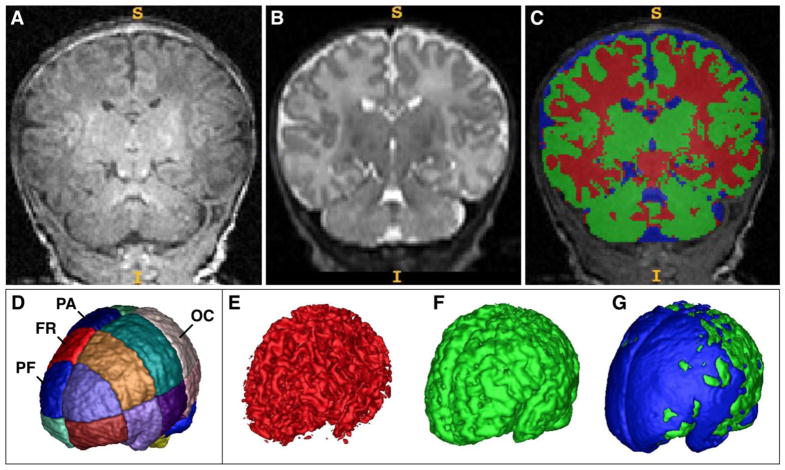
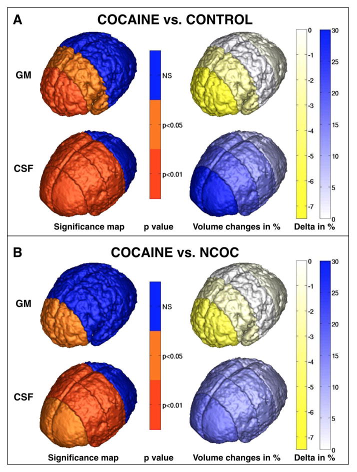
Prenatal cocaine exposure (PCE) is related to subtle deficits in cognitive and behavioral function in infancy, childhood and adolescence. Very little is known about the effects of in utero PCE on early brain development that may contribute to these impairments. The purpose of this study was to examine brain structural differences in infants with and without PCE. We conducted MRI scans of newborns (mean age = 5 weeks) to determine cocaine's impact on early brain structural development. Subjects were three groups of infants: 33 with PCE co-morbid with other drugs, 46 drug-free controls and 40 with prenatal exposure to other drugs (nicotine, alcohol, marijuana, opiates, SSRIs) but without cocaine. Infants with PCE exhibited lesser total gray matter (GM) volume and greater total cerebral spinal fluid (CSF) volume compared with controls and infants with non-cocaine drug exposure. Analysis of regional volumes revealed that whole brain GM differences were driven primarily by lesser GM in prefrontal and frontal brain regions in infants with PCE, while more posterior regions (parietal, occipital) did not differ across groups. Greater CSF volumes in PCE infants were present in prefrontal, frontal and parietal but not occipital regions. Greatest differences (GM reduction, CSF enlargement) in PCE infants were observed in dorsal prefrontal cortex. Results suggest that PCE is associated with structural deficits in neonatal cortical gray matter, specifically in prefrontal and frontal regions involved in executive function and inhibitory control. Longitudinal study is required to determine whether these early differences persist and contribute to deficits in cognitive functions and enhanced risk for drug abuse seen at school age and in later life.
Keywords: CSF enlargement; Cortical gray matter; Infant brain development; Magnetic resonance imaging; Prenatal cocaine; Prenatal substance abuse.
Copyright © 2014 Elsevier Inc. All rights reserved.
Figures
References
-
- Avants BB, Hurt H, et al. Effects of heavy in utero cocaine exposure on adolescent caudate morphology. Pediatr Neurol. 2007;37(4):275–279. - PubMed
-
- Bandstra ES, Morrow CE, et al. Longitudinal investigation of task persistence and sustained attention in children with prenatal cocaine exposure. Neurotoxicol Teratol. 2001;23(6):545–559. - PubMed
Publication types
MeSH terms
Substances
Grants and funding
- M01 RR000046/RR/NCRR NIH HHS/United States
- 1P01DA022446-01A1/DA/NIDA NIH HHS/United States
- U54EB005149/EB/NIBIB NIH HHS/United States
- P50 MH064065/MH/NIMH NIH HHS/United States
- R01MH070890/MH/NIMH NIH HHS/United States
- U54 HD079124/HD/NICHD NIH HHS/United States
- R01 MH070890/MH/NIMH NIH HHS/United States
- K01 DA019949/DA/NIDA NIH HHS/United States
- P01 DA022446/DA/NIDA NIH HHS/United States
- U54 EB005149/EB/NIBIB NIH HHS/United States
- K01DA019949-01A1/DA/NIDA NIH HHS/United States
- MH064065/MH/NIMH NIH HHS/United States
LinkOut - more resources
Full Text Sources
Other Literature Sources
