. 2014 Jun 20:6:132.
doi: 10.3389/fnagi.2014.00132. eCollection 2014.

Selective estrogen receptor modulators regulate reactive microglia after penetrating brain injury

Affiliations

Selective estrogen receptor modulators regulate reactive microglia after penetrating brain injury

George E Barreto et al. Front Aging Neurosci. .

Abstract

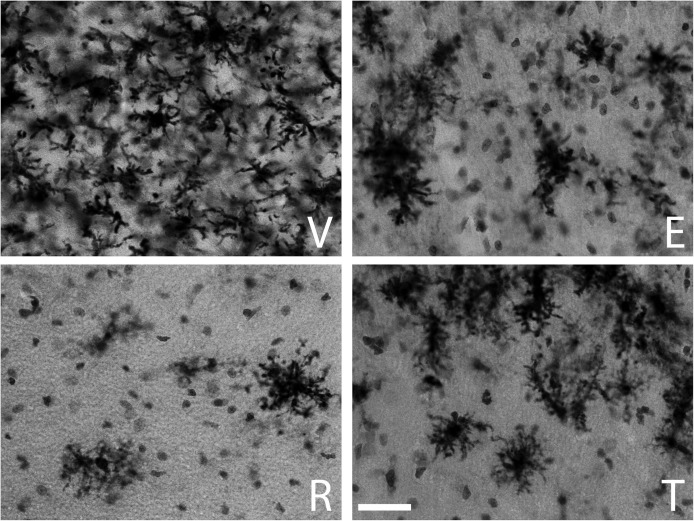
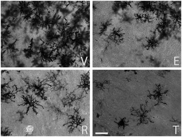
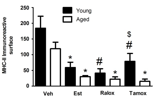
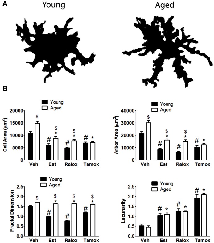
Following brain injury, microglia assume a reactive-like state and secrete pro-inflammatory molecules that can potentiate damage. A therapeutic strategy that may limit microgliosis is of potential interest. In this context, selective estrogen receptor modulators, such as raloxifene and tamoxifen, are known to reduce microglia activation induced by neuroinflammatory stimuli in young animals. In the present study, we have assessed whether raloxifene and tamoxifen are able to affect microglia activation after brain injury in young and aged animals in time points relevant to clinics, which is hours after brain trauma. Volume fraction of MHC-II(+) microglia was estimated according to the point-counting method of Weibel within a distance of 350 μm from the lateral border of the wound, and cellular morphology was measured by fractal analysis. Two groups of animals were studied: (1) young rats, ovariectomized at 2 months of age; and (2) aged rats, ovariectomized at 18 months of age. Fifteen days after ovariectomy animals received a stab wound brain injury and the treatment with estrogenic compounds. Our findings indicate that raloxifene and tamoxifen reduced microglia activation in both young and aged animals. Although the volume fraction of reactive microglia was found lower in aged animals, this was accompanied by important changes in cell morphology, where aged microglia assume a bushier and hyperplasic aspect when compared to young microglia. These data suggest that early regulation of microglia activation provides a mechanism by which selective estrogen receptors modulators (SERMs) may exert a neuroprotective effect in the setting of a brain trauma.

Keywords: brain injury; microglia; neuroprotection; raloxifene; tamoxifen.


Figures

Figure 1
Figure 1
MHC-II immunoreactive microglia in the CA1 stratum radiatum at a distance of approximately 100–200 µm from the lateral border of the wound in young animals. The panels illustrate representative examples from ovariectomized young rats after administration of (V) vehicle, (E) estradiol, (R) raloxifene or (T) tamoxifen. All figures are at the same magnification. Scale bar, 50 um.
Figure 2
Figure 2
MHC-II immunoreactive microglia in the CA1 stratum radiatum at a distance of approximately 100–200 µm from the lateral border of the wound in aged animals. The panels illustrate representative examples from ovariectomized aged rats after administration of (V) vehicle, (E) estradiol, (R) raloxifene or (T) tamoxifen. All figures are at the same magnification. Scale bar, 50 um.
Figure 3
Figure 3
Volume fraction of MHC-II immunoreactive microglia within a distance of 350 µm from the lateral border of the wound in young and aged animals injected with vehicle (Veh) n = 6 (young) and 5 (aged), estradiol (Est) n = 4 (young and aged), raloxifene (Ralox) n = 6 (young) and 4 (aged), or tamoxifen (Tamox) n = 5 (young) and 4 (aged). Data are represented as means +/− SEM. * Significant difference (P < 0.05) vs. vehicle values. * P < 0.05 vs. vehicle (young); # P < 0.05 vs. vehicle (aged); $ P < 0.05 young vs. aged treated with same SERM.
Figure 4
Figure 4
(A) The upper panel indicates age differences on microglia morphology with aging in animals treated with vehicle. Fractal analysis shows that microglia reactivity is characterized by changes in both cell body and processes. (B) Cell shape is hypertrophied and bushier in control cells (vehicle, V) compared to those cells from animals treated with estradiol (Est), raloxifene (Ralox) or tamoxifen (Tamox). * P < 0.05 vs. vehicle (young); # P < 0.05 vs. vehicle (aged); $ P < 0.05 young vs. aged treated with same SERM.

References

    1. Adelson J. D., Barreto G. E., Xu L., Kim T., Brott B. K., Ouyang Y. B., et al. (2012). Neuroprotection from stroke in the absence of MHCI or PirB. Neuron 73, 1100–1107 10.1016/j.neuron.2012.01.020 - DOI - PMC - PubMed
    1. Aloisi F. (2001). Immune function of microglia. Glia 36, 165–179 10.1002/glia.1106 - DOI - PubMed
    1. Arevalo M. A., Diz-Chaves Y., Santos-Galindo M., Bellini M. J., Garcia-Segura L. M. (2012). Selective oestrogen receptor modulators decrease the inflammatory response of glial cells. J. Neuroendocrinol. 24, 183–190 10.1111/j.1365-2826.2011.02156.x - DOI - PubMed
    1. Baker A. E., Brautigam V. M., Watters J. J. (2004). Estrogen modulates microglial inflammatory mediator production via interactions with estrogen receptor beta. Endocrinology 145, 5021–5032 10.1210/en.2004-0619 - DOI - PubMed
    1. Barreto G. E., Gonzalez J., Torres Y., Morales L. (2011). Astrocytic-neuronal crosstalk: implications for neuroprotection from brain injury. Neurosci. Res. 71, 107–113 10.1016/j.neures.2011.06.004 - DOI - PubMed