. 2014 Jun 1;1(6):379-389.
doi: 10.1002/acn3.64.

Lysosomal abnormalities in hereditary spastic paraplegia types SPG15 and SPG11

Affiliations

Lysosomal abnormalities in hereditary spastic paraplegia types SPG15 and SPG11

Benoît Renvoisé et al. Ann Clin Transl Neurol. .

Abstract

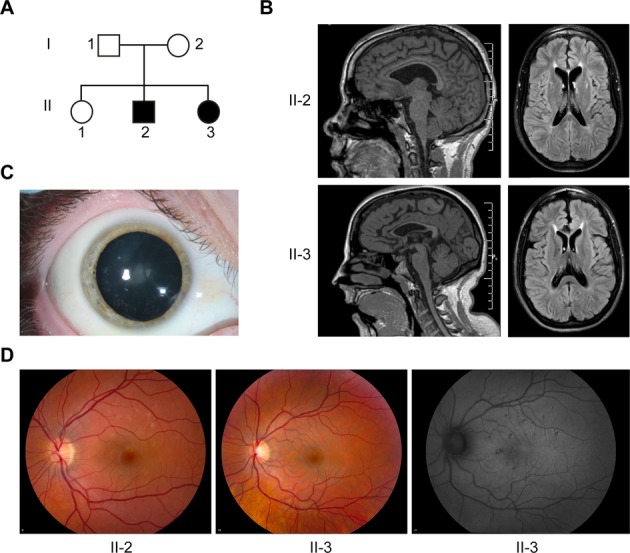
Objective: Hereditary spastic paraplegias (HSPs) are among the most genetically diverse inherited neurological disorders, with over 70 disease loci identified (SPG1-71) to date. SPG15 and SPG11 are clinically similar, autosomal recessive disorders characterized by progressive spastic paraplegia along with thin corpus callosum, white matter abnormalities, cognitive impairment, and ophthalmologic abnormalities. Furthermore, both have been linked to early-onset parkinsonism.

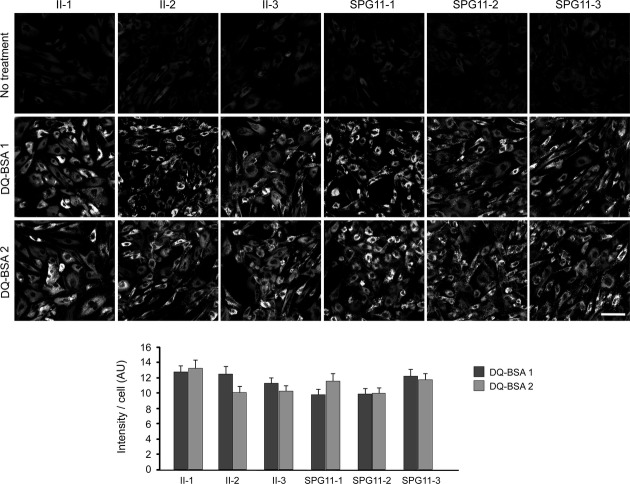
Methods: We describe two new cases of SPG15 and investigate cellular changes in SPG15 and SPG11 patient-derived fibroblasts, seeking to identify shared pathogenic themes. Cells were evaluated for any abnormalities in cell division, DNA repair, endoplasmic reticulum, endosomes, and lysosomes.

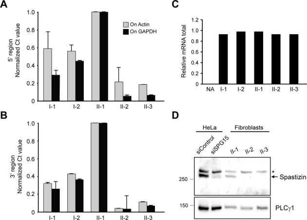
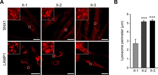
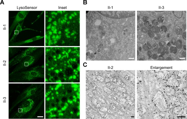
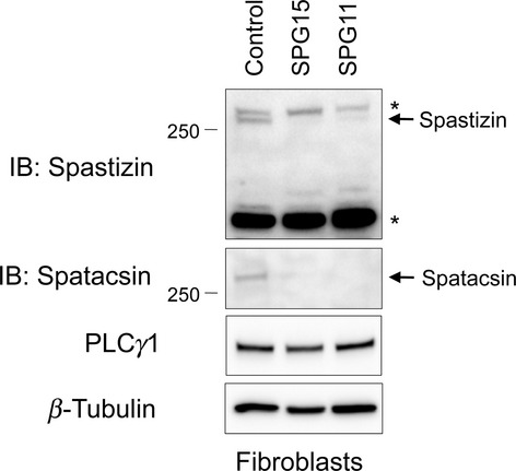
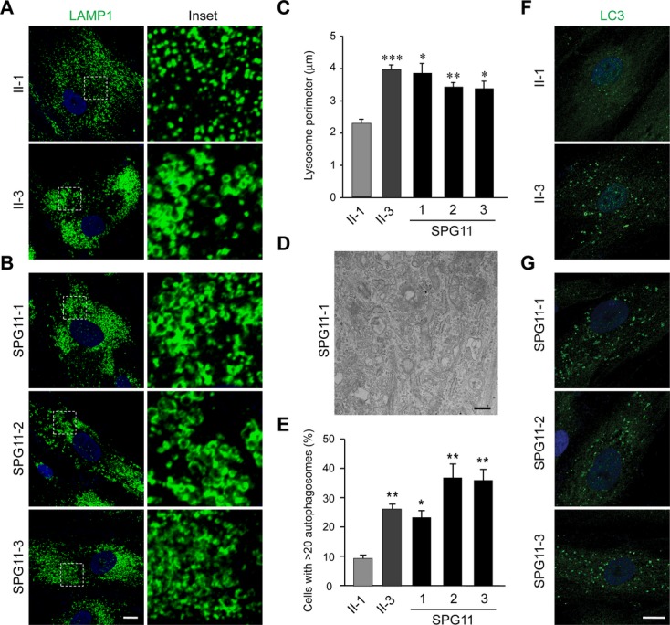
Results: Fibroblasts prepared from patients with SPG15 have selective enlargement of LAMP1-positive structures, and they consistently exhibited abnormal lysosomal storage by electron microscopy. A similar enlargement of LAMP1-positive structures was also observed in cells from multiple SPG11 patients, though prominent abnormal lysosomal storage was not evident. The stabilities of the SPG15 protein spastizin/ZFYVE26 and the SPG11 protein spatacsin were interdependent.

Interpretation: Emerging studies implicating these two proteins in interactions with the late endosomal/lysosomal adaptor protein complex AP-5 are consistent with shared abnormalities in lysosomes, supporting a converging mechanism for these two disorders. Recent work with Zfyve26-/- mice revealed a similar phenotype to human SPG15, and cells in these mice had endolysosomal abnormalities. SPG15 and SPG11 are particularly notable among HSPs because they can also present with juvenile parkinsonism, and this lysosomal trafficking or storage defect may be relevant for other forms of parkinsonism associated with lysosomal dysfunction.


Figures

Figure 1
Figure 1
Clinical and imaging features of SPG15. (A) Pedigree of SPG15 family. Square: male; circle: female; solid symbol: affected individual. (B) T1-weighted sagittal MRI images (left) show enlarged ventricles and TCC in the SPG15 affected siblings. Axial fluid attenuation inversion recovery images (right) show increased signal particularly in the region of the forceps minor, just anterior to the rostral-most portion of the lateral ventricles. (C) Small, white, peripheral cortical lens opacities are evident in SPG15 patient II-3 (right eye shown). (D) Yellowish flecks are notable in the retinas of SPG15 patients, particularly in II-2; these were more apparent by autofluorescence in II-3 (right panel).
Figure 2
Figure 2
The SPG15 protein spastizin is absent in SPG15 patient fibroblasts. (A–C) Quantitative PCR analysis of spastizin mRNA using primers targeting both 5′ (A) and 3′ (B) ends. Actin and GADPH were used as standards. Graphs show mean ± SD. Relative total mRNA levels are shown in (C). Similar results were obtained in at least two additional independent experiments. NA, negative control. (D) Immunoblot analysis of spastizin in HeLa cells transfected with control or spastizin-specific siRNAs as well as cultured fibroblasts from the indicated subjects. An asterisk (*) identifies a cross-reacting protein. PLCγ1 levels were monitored as a control for protein loading. Migrations of protein standards (in kDa) are at the left.
Figure 3
Figure 3
SPG15 fibroblast cells exhibit prominently enlarged lysosomes. (A) Immunostaining of LAMP1 and SNX1 (both in red) proteins in the indicated fibroblast cell lines. Boxed areas are enlarged in the insets. Scale bars: 20 μm. (B) Quantification of lysosome circumference from subject-derived cells, mean ± SEM (n = 3 independent experiments, each evaluating at least 250 lysosomes). ***P < 0.001; unpaired Student's t test.
Figure 4
Figure 4
SPG15 fibroblasts harbor numerous multilamellar zebra bodies. (A) Confocal microscopy analysis of the indicated fibroblast lines stained with LysoSensor Green. Boxed areas in the left panels are enlarged at the right. Scale bar: 10 μm. (B) EM reveals numerous membranous multilamellar zebra bodies throughout the cytoplasm of fibroblasts cultured from subject II-3 (shown) and II-2 (not shown), who have SPG15, but not in cells from an unaffected sister (I-1). Scale bars: 500 nm. (C) Immunogold-EM shows LAMP1 immunoreactivity rimming storage-filled structures. These structures appear different than in B because here cells were permeabilized with the detergent saponin, altering the membranes, and the OsO4 concentration was much lower, decreasing membrane contrast. Scale bars: 500 nm.
Figure 5
Figure 5
Both spastizin and spatacsin proteins are depleted from SPG15- and SPG11-patient cell lines. Cell extracts prepared from SPG15 (II-2) and SPG11 patient cell lines were immunoblotted (IB) as shown. PLCγ1 and β-tubulin levels were monitored as controls for protein loading. Asterisks (*) identify cross-reacting proteins. Migrations of protein standards (in kDa) are at the left.
Figure 6
Figure 6
Fibroblasts from SPG11 patients also exhibit enlarged LAMP1-positive structures. (A and B) Confocal microscopy analysis of the fibroblast cell lines stained with LAMP1 antibodies: (A) control and SPG15 fibroblasts; (B) SPG11 fibroblasts. Insets in the left panels are enlarged at the right. Nuclei are stained with DAPI. (C) Quantification of lysosome circumference from subject-derived cells, mean ± SEM (n = 3 independent experiments, each evaluating at least 250 lysosomes). (D) EM of SPG11 fibroblasts. Scale bar: 500 nm. (E–G) LC3-positive autophagosomes were counted in the indicated fibroblast lines (mean ± SD, n = 3 independent experiments, each evaluating at least 200 cells). DAPI staining identifies the nucleus. Scale bar: 10 μm. ***P < 0.001; **P < 0.01; *P < 0.05; paired Student's t test.
Figure 7
Figure 7
Lysosomes of SPG11 and SPG15 patient cells appear enzymatically functional. Control (II-1) and patient fibroblast cells labeled by DQ-BSA-red were imaged. The intensities of manually determined areas for each single cell were measured using ImageJ measurement tools. All cell lines were analyzed in duplicate for DQ-BSA treatment. The graph shows mean ± SEM (n = 30). AU, arbitrary unit. Scale bar: 100 μm.

References

    1. Blackstone C. Cellular pathways of hereditary spastic paraplegia. Annu Rev Neurosci. 2012;35:25–47. - PMC - PubMed
    1. Finsterer J, Löscher W, Quasthoff S, et al. Hereditary spastic paraplegias with autosomal dominant, recessive, X-linked, or maternal trait of inheritance. J Neurol Sci. 2012;318:1–18. - PubMed
    1. Park SH, Zhu P-P, Parker RL, Blackstone C. Hereditary spastic paraplegia proteins REEP1, spastin, and atlastin-1 coordinate microtubule interactions with the tubular ER network. J Clin Invest. 2010;120:1097–1110. - PMC - PubMed
    1. Blackstone C, O'Kane CJ, Reid E. Hereditary spastic paraplegias: membrane traffic and the motor pathway. Nat Rev Neurosci. 2011;12:31–42. - PMC - PubMed
    1. Novarino G, Fenstermaker AG, Zaki MS, et al. Exome sequencing links corticospinal motor neuron disease to common neurodegenerative disorders. Science. 2014;343:506–511. - PMC - PubMed