

Developmental lead exposure alters synaptogenesis through inhibiting canonical Wnt pathway in vivo and in vitro
- PMID: 24999626
- PMCID: PMC4084981
- DOI: 10.1371/journal.pone.0101894
Developmental lead exposure alters synaptogenesis through inhibiting canonical Wnt pathway in vivo and in vitro
Abstract
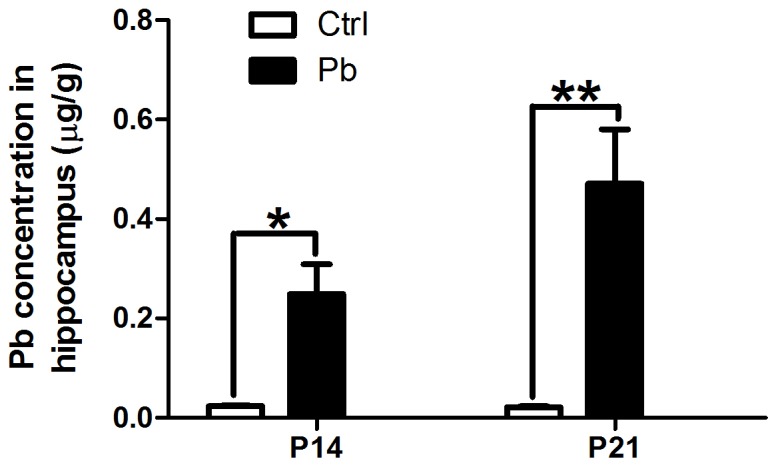
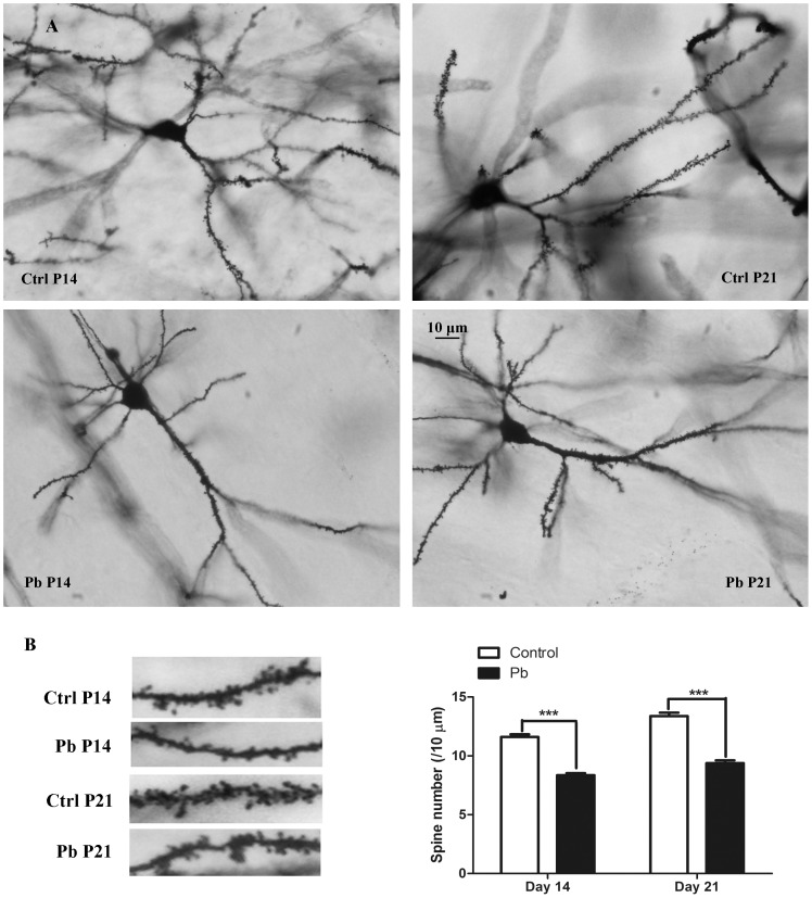
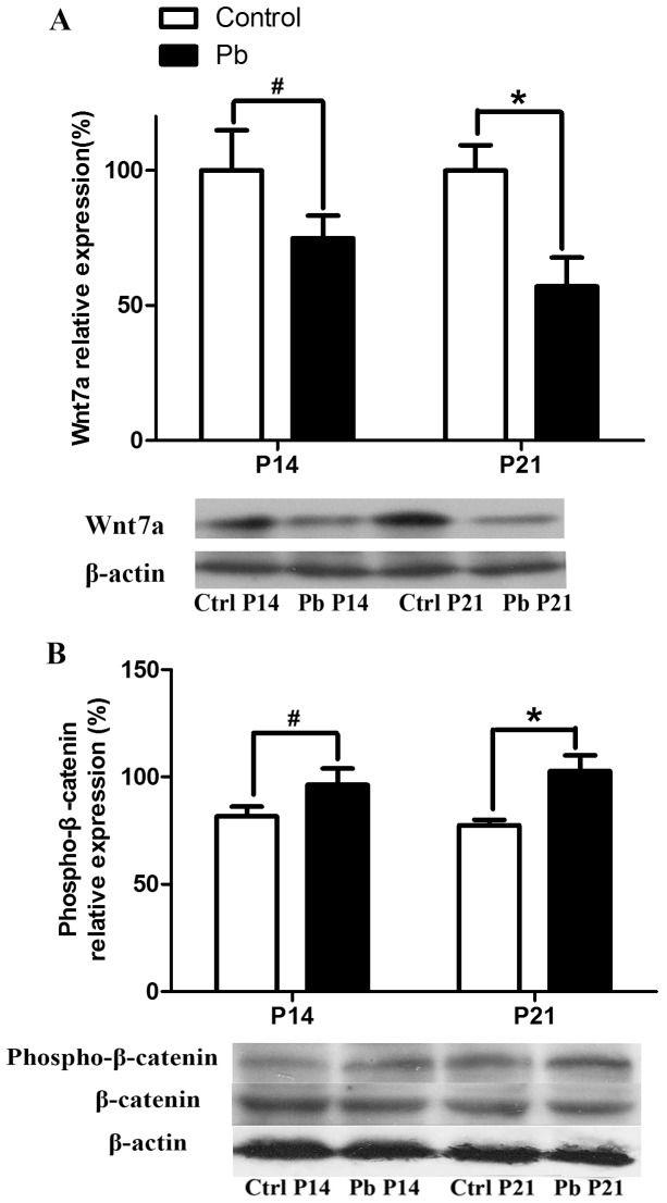
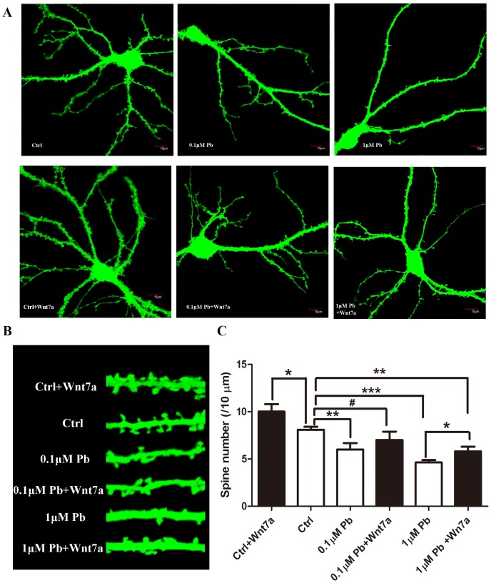
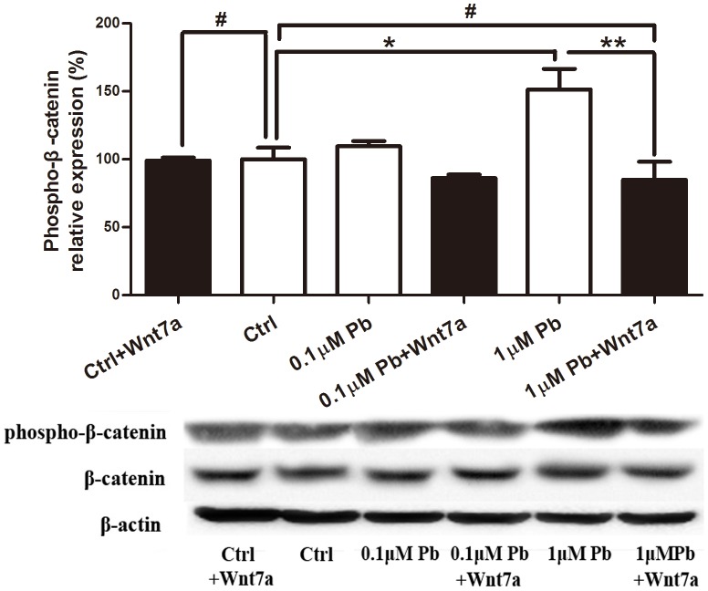
Lead (Pb) exposure has been implicated in the impairment of synaptic plasticity in the developing hippocampus, but the mechanism remains unclear. Here, we investigated whether developmental lead exposure affects the dendritic spine formation through Wnt signaling pathway in vivo and in vitro. Sprague-Dawley rats were exposed to lead throughout the lactation period and Golgi-Cox staining method was used to examine the spine density of pyramidal neurons in the hippocampal CA1 area of rats. We found that lead exposure significantly decreased the spine density in both 14 and 21 days-old pups, accompanied by a significant age-dependent decline of the Wnt7a expression and stability of its downstream protein (β-catenin). Furthermore, in cultured hippocampal neurons, lead (0.1 and 1 µM lead acetate) significantly decreased the spine density in a dose-dependent manner. Exogenous Wnt7a application attenuated the decrease of spine density and increased the stability of the downstream molecules in Wnt signaling pathway. Together, our results suggest that lead has a negative impact on spine outgrowth in the developing hippocampus through altering the canonical Wnt pathway.
Conflict of interest statement
Figures
Similar articles
-
Low-level Gestational Lead Exposure Alters Dendritic Spine Plasticity in the Hippocampus and Reduces Learning and Memory in Rats.Sci Rep. 2018 Feb 23;8(1):3533. doi: 10.1038/s41598-018-21521-8. Sci Rep. 2018. PMID: 29476096 Free PMC article.
-
β-Asarone Rescues Pb-Induced Impairments of Spatial Memory and Synaptogenesis in Rats.PLoS One. 2016 Dec 9;11(12):e0167401. doi: 10.1371/journal.pone.0167401. eCollection 2016. PLoS One. 2016. PMID: 27936013 Free PMC article.
-
Wnt7a signaling promotes dendritic spine growth and synaptic strength through Ca²⁺/Calmodulin-dependent protein kinase II.Proc Natl Acad Sci U S A. 2011 Jun 28;108(26):10732-7. doi: 10.1073/pnas.1018132108. Epub 2011 Jun 13. Proc Natl Acad Sci U S A. 2011. PMID: 21670302 Free PMC article.
-
Early developmental bisphenol-A exposure sex-independently impairs spatial memory by remodeling hippocampal dendritic architecture and synaptic transmission in rats.Sci Rep. 2016 Aug 31;6:32492. doi: 10.1038/srep32492. Sci Rep. 2016. PMID: 27578147 Free PMC article.
-
[Involvement of Wnt signaling in hippocampal plasticity].Ross Fiziol Zh Im I M Sechenova. 2012 Dec;98(12):1460-70. Ross Fiziol Zh Im I M Sechenova. 2012. PMID: 23461192 Review. Russian.
Cited by
-
Exposure of metal toxicity in Alzheimer's disease: An extensive review.Front Pharmacol. 2022 Aug 29;13:903099. doi: 10.3389/fphar.2022.903099. eCollection 2022. Front Pharmacol. 2022. PMID: 36105221 Free PMC article. Review.
-
Strain specific effects of low level lead exposure on associative learning and memory in rats.Neurotoxicology. 2017 Sep;62:186-191. doi: 10.1016/j.neuro.2017.07.006. Epub 2017 Jul 16. Neurotoxicology. 2017. PMID: 28720388 Free PMC article.
-
Effects of Gastrodin against Lead-Induced Brain Injury in Mice Associated with the Wnt/Nrf2 Pathway.Nutrients. 2020 Jun 17;12(6):1805. doi: 10.3390/nu12061805. Nutrients. 2020. PMID: 32560430 Free PMC article.
-
Mechanisms of divalent metal toxicity in affective disorders.Toxicology. 2016 Jan 2;339:58-72. doi: 10.1016/j.tox.2015.11.001. Epub 2015 Nov 10. Toxicology. 2016. PMID: 26551072 Free PMC article. Review.
-
Cerebral Vascular Toxicity after Developmental Exposure to Arsenic (As) and Lead (Pb) Mixtures.Toxics. 2024 Aug 24;12(9):624. doi: 10.3390/toxics12090624. Toxics. 2024. PMID: 39330552 Free PMC article.
References
-
- Meng XM, Zhu DM, Ruan DY, She JQ, Luo L (2005) Effects of chronic lead exposure on 1H MRS of hippocampus and frontal lobes in children. Neurology 64: 1644–1647. - PubMed
Publication types
MeSH terms
Substances
LinkOut - more resources
Full Text Sources
Other Literature Sources
Miscellaneous
