Review
. 2014 Oct:30:74-82.
doi: 10.1016/j.ceb.2014.06.005. Epub 2014 Jul 5.

Directed migration of mesenchymal cells: where signaling and the cytoskeleton meet

Affiliations
Review

Directed migration of mesenchymal cells: where signaling and the cytoskeleton meet

James E Bear et al. Curr Opin Cell Biol. 2014 Oct.

Abstract

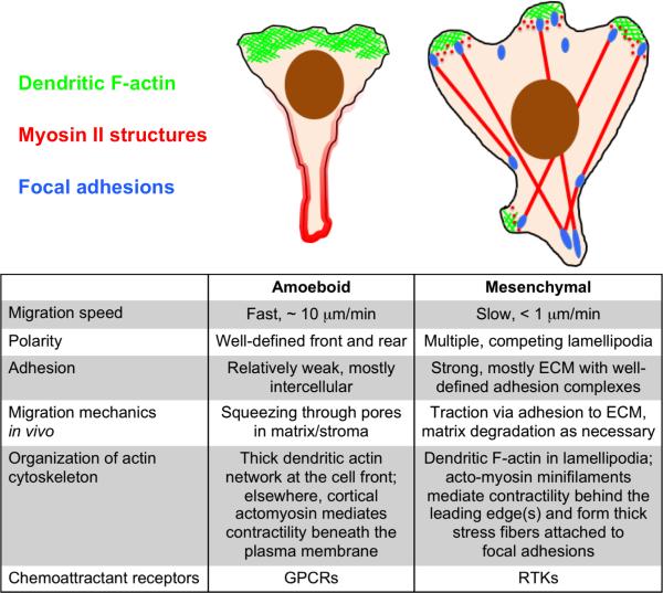
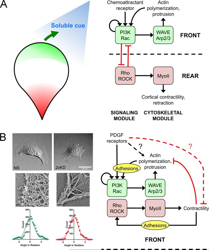
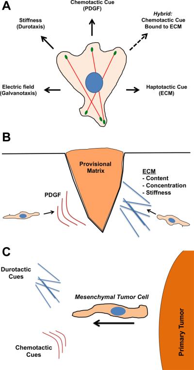
Cell migration directed by spatial cues, or taxis, is a primary mechanism for orchestrating concerted and collective cell movements during development, wound repair, and immune responses. Compared with the classic example of amoeboid chemotaxis, in which fast-moving cells such as neutrophils are directed by gradients of soluble factors, directed migration of slow-moving mesenchymal cells such as fibroblasts is poorly understood. Mesenchymal cells possess a distinctive organization of the actin cytoskeleton and associated adhesion complexes as its primary mechanical system, generating the asymmetric forces required for locomotion without strong polarization. The emerging hypothesis is that the molecular underpinnings of mesenchymal taxis involve distinct signaling pathways and diverse requirements for regulation.


Figures

Figure 1
Figure 1. Mesenchymal vs. amoeboid motility and chemotaxis
The illustrations and table compare the structural and dynamic features of mesenchymal migration to those of amoeboid cells such as neutrophils and lymphocytes.
Figure 2
Figure 2. Clarifying the role of the PI3K/Rac/WAVE/Arp2/3 circuit in mesenchymal cells
(A) In the conventional model of gradient sensing in amoeboid cells, PI3K and Rac are engaged in a signaling module that controls Arp2/3-mediated actin polymerization at the front of the cell. Cells establish and maintain polarity through positive feedback in this circuit, combined with its functional incompatibility with Rho signaling and active Myosin II (MyoII) at the cell rear. An external cue simply introduces a bias of the Arp2/3 circuit towards the left or right of the migration axis. (B) The alternative model of mesenchymal chemotaxis is spurred by the observation that depletion of Arp2/3 complex in fibroblasts results in loss of dendritic F-actin arrays associated with lamellipodia but does not affect chemotactic fidelity. Panels at left adapted from Wu et al. [55••] (Copyright 2012 Elsevier Inc., used with permission). In this model, the Arp2/3 circuit follows the cue of an as yet uncharacterized gradient sensing mechanism and is important for agile cell movement during chemotaxis as well as random migration. This functional distinction is consistent with the relative lack of polarization in mesenchymal cells, in which Arp2/3 and myosin modules are neighbors, and the role of adhesion complexes in activating them to elicit protrusion and retraction of lamellipodia, respectively.
Figure 3
Figure 3. Directed migration cues for mesenchymal cells
(A) Diagram illustrating the diverse types of directional cues that mesenchymal cells respond to. Of note is the hybrid cue where chemotactic cues (e.g., growth factors) are bound to ECM scaffolds. (B) During cutaneous wound healing, fibroblasts (prototypical mesenchymal cells) respond to both PDGF (chemotaxis) and ECM cues (haptotaxis/durotaxis). (C) Likewise, mesenchymal tumor cells emerging from primary tumors sense multiple directional cues.

References

    1. Singer AJ, Clark RAF. Mechanisms of disease: cutaneous wound healing. N Engl J Med. 1999;341:738–746. - PubMed
    1. Deuel TF, Kawahara RS, Mustoe TA, Pierce GF. Growth factors and wound healing: platelet-derived growth factor as a model cytokine. Annu Rev Med. 1991;42:567–584. - PubMed
    1. Ataliotis P, Mercola M. Distribution and functions of platelet-derived growth factors and their receptors during embryogenesis. Intl Rev Cytol. 1997;172:95–127. - PubMed
    1. Edelberg JM, Cai D, Xaymardan M. Translation of PDGF cardioprotective pathways. Cardiovasc Toxicol. 2003;3:27–35. - PubMed
    1. Leask A. Potential therapeutic targets for cardiac fibrosis: TGFb, angiotensin, endothelin, CCN2, and PDGF, partners in fibroblast activation. Circ Res. 2010;106:1675–1680. - PubMed

Publication types

LinkOut - more resources