. 2014 Jul 7;9(7):e99409.
doi: 10.1371/journal.pone.0099409. eCollection 2014.

Clinical validation of an ultra high-throughput spiral microfluidics for the detection and enrichment of viable circulating tumor cells

Affiliations

Clinical validation of an ultra high-throughput spiral microfluidics for the detection and enrichment of viable circulating tumor cells

Bee Luan Khoo et al. PLoS One. .

Erratum in

  • PLoS One. 2014;9(10):e111296

Abstract

Background: Circulating tumor cells (CTCs) are cancer cells that can be isolated via liquid biopsy from blood and can be phenotypically and genetically characterized to provide critical information for guiding cancer treatment. Current analysis of CTCs is hindered by the throughput, selectivity and specificity of devices or assays used in CTC detection and isolation.

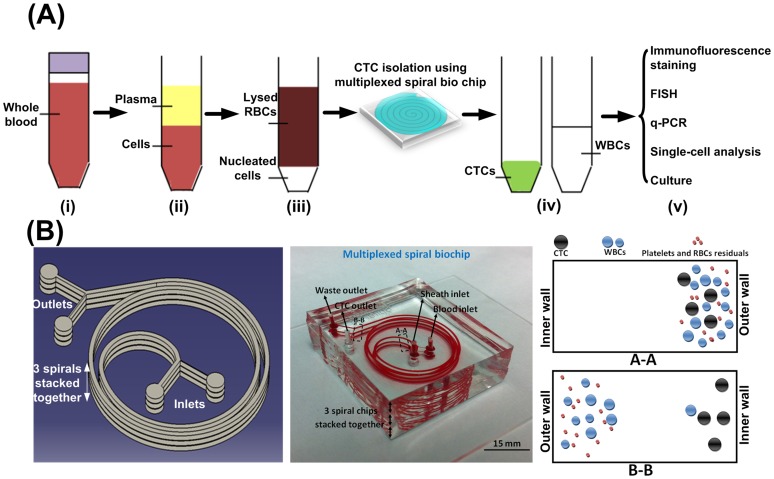
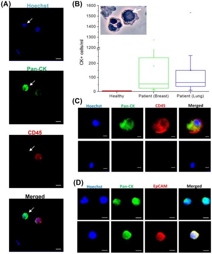
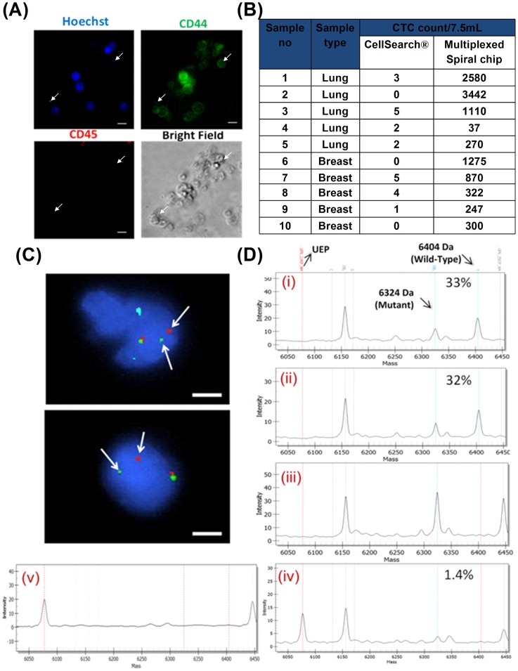
Methodology/principal findings: Here, we enriched and characterized putative CTCs from blood samples of patients with both advanced stage metastatic breast and lung cancers using a novel multiplexed spiral microfluidic chip. This system detected putative CTCs under high sensitivity (100%, n = 56) (Breast cancer samples: 12-1275 CTCs/ml; Lung cancer samples: 10-1535 CTCs/ml) rapidly from clinically relevant blood volumes (7.5 ml under 5 min). Blood samples were completely separated into plasma, CTCs and PBMCs components and each fraction were characterized with immunophenotyping (Pan-cytokeratin/CD45, CD44/CD24, EpCAM), fluorescence in-situ hybridization (FISH) (EML4-ALK) or targeted somatic mutation analysis. We used an ultra-sensitive mass spectrometry based system to highlight the presence of an EGFR-activating mutation in both isolated CTCs and plasma cell-free DNA (cf-DNA), and demonstrate concordance with the original tumor-biopsy samples.

Conclusions/significance: We have clinically validated our multiplexed microfluidic chip for the ultra high-throughput, low-cost and label-free enrichment of CTCs. Retrieved cells were unlabeled and viable, enabling potential propagation and real-time downstream analysis using next generation sequencing (NGS) or proteomic analysis.


Conflict of interest statement

Competing Interests: AASB is an employee of Clearbridge BioMedics Pte Ltd which commercialized the technology described here. AASB, JH and CTL, along with others, also have filed a patent application on the technology. Pending patent: Microfluidics Sorter for Cell Detection and Isolation and number PCT/US2011/027276. A product based on this technology is currently being developed by Clearbridge Biomedics Pte Ltd. This does not alter the authors' adherence to all the PLOS ONE policies on sharing data and materials.

Figures

Figure 1
Figure 1. Overview of sample preparation and processing procedures.
(A) Sample processing workflow showing different steps of enrichment and identification. (i) The blood sample is collected; (ii) Plasma is separated using standard centrifugation (1500×g for 10 min) and stored at −80 degree Celsius for DNA analysis. (iii) RBCs are lysed using ammonium chloride and (iv) sample is processed through multiplexed spiral chip within 10 min. (v) The isolated CTCs are available for immunostaining using standard markers or FISH (fluorescence in situ hybridization). DNA or RNA can be extracted from the CTCs and subjected to next-generation sequencing and q-PCR. Viable cells can be released and propagated in cell culture for various applications including cancer stem cell (CSC) study or drug discovery. (B) Illustration of the design of a multiplexed device (left) and optical image of an actual multiplexed spiral microfluidic device (middle) for capturing CTCs with two inlets and two outlets. Blood sample and sheath fluid are pumped through the device using two separate syringe pumps. Under the influence of inertial lift and Dean drag forces in the fluid flow, CTCs focus near microchannel inner wall (Region A-A) while WBCs and Platelets goes through one Dean cycle and migrate back towards the outer wall (Region B-B), thus achieving separation.
Figure 2
Figure 2. Enumeration of CTC from cancer patients.
(A) Immunofluorescence staining of isolated CTCs. CTCs (marked by white arrow) were identified by the following criteria: Hoechst+, pan-CK+ and CD45-. Scale bar: 20 µm (B) Box plot summary indicating the range of CK+cells/ml recovered from the sample outlet for blood samples extracted from healthy volunteers, as wells as breast and lung cancer patients. The box plot presents the median, lower and upper quartiles (25th,75th percentiles). Data points that lie outside the 10th and 90th percentiles are shown as outliers (Anova, p<0.001). Encapsulated image of PAP stained isolated cells shows a large CTC with high nucleus to cytoplasmic (N/C) ratio (labeled with white arrow). (C) Staining of CTC for pan-CK and CD45. Upper panel depicts a representative image of cells which were double positive (CK+/CD45+); while lower panel shows a double negative (CK-/CD45-) cell. Scale bar: 20 µm (D) Staining of CTC for pan-cytokeratin and EpCAM. Scale bar: 20 µm.
Figure 3
Figure 3. Downstream analysis of enriched CTCs.
(A) CTC viability demonstrated by attachment to 2D Geltrex (Invitrogen)-coated substrate (72 hr after seeding). Isolated CTC were enriched for CD44. No cells were stained for CD45, indicating the absence of WBCs which did not adhere to substrate and were removed after washing with 1X PBS. Some CD44+cells were not stained for Hoechst (white arrows). Scale bar: 20 µm (B) Comparison of CTC isolation and recovery with CellSearch system. (C) Molecular FISH analysis on enriched CTCs of a patient with NSCLC. Cells were stained using Vysis ALK Break Apart FISH probe and counterstained with DAPI. The red and green signals demonstrated a distinct separation of the original fusion signal (arrows), indicating a rearrangement in the 2p23 ALK-gene locus. Scale bar: 16 µm. (D) MassArray spectra for a patient with NSCLC harboring EGFR L747_P753>S. Trace from FFPE, plasma and pooled CTCs illustrated. Percentage indicates calculated proportion of mutant allele against wild type allele (UEP: Unextended primer). (i) iPlex bi-allelic spectra on FFPE sample (33% mutant frequency), (ii) iPlex bi-allelic spectrum on plasma sample (32% mutant frequency), (iii) SABER mutant specific spectrum on plasma sample (Positive – high frequency), (iv) SABER mutant specific spectrum on CTCs (Positive – low frequency (n = 3/94), estimated mutant frequency of 1.4%) and (v) Representative iPlex & SABER (shown) spectrum on no-template control sample (Negative).

References

    1. Pantel K, Brakenhoff RH, Brandt B (2008) Detection, clinical relevance and specific biological properties of disseminating tumour cells. Nature Reviews Cancer 8: 329–340. - PubMed
    1. Majid EW, Lim CT (2013) Microfluidic Platforms for Human Disease Cell Mechanics Studies. Materiomics: Multiscale Mechanics of Biological Materials and Structures: Springer. pp. 107–119.
    1. He W, Wang H, Hartmann LC, Cheng JX, Low PS (2007) In vivo quantitation of rare circulating tumor cells by multiphoton intravital flow cytometry. Proceedings of the National Academy of Sciences 104: 11760. - PMC - PubMed
    1. Allan AL, Vantyghem SA, Tuck AB, Chambers AF, Chin-Yee IH, et al. (2005) Detection and quantification of circulating tumor cells in mouse models of human breast cancer using immunomagnetic enrichment and multiparameter flow cytometry. Cytometry Part A 65: 4–14. - PubMed
    1. Gertler R, Rosenberg R, Fuehrer K, Dahm M, Nekarda H, et al. (2003) Detection of circulating tumor cells in blood using an optimized density gradient centrifugation. Recent Results in Cancer Research 162: 149–156. - PubMed

Publication types

MeSH terms