. 2014 Jul 8:15:27.
doi: 10.1186/1471-2121-15-27.

Distinct genetic programs guide Drosophila circular and longitudinal visceral myoblast fusion

Affiliations

Distinct genetic programs guide Drosophila circular and longitudinal visceral myoblast fusion

Anja Rudolf et al. BMC Cell Biol. .

Abstract

Background: The visceral musculature of Drosophila larvae comprises circular visceral muscles tightly interwoven with longitudinal visceral muscles. During myogenesis, the circular muscles arise by one-to-one fusion of a circular visceral founder cell (FC) with a visceral fusion-competent myoblast (FCM) from the trunk visceral mesoderm, and longitudinal muscles arise from FCs of the caudal visceral mesoderm. Longitudinal FCs migrate anteriorly under guidance of fibroblast growth factors during embryogenesis; it is proposed that they fuse with FCMs from the trunk visceral mesoderm to give rise to syncytia containing up to six nuclei.

Results: Using fluorescence in situ hybridization and immunochemical analyses, we investigated whether these fusion events during migration use the same molecular repertoire and cellular components as fusion-restricted myogenic adhesive structure (FuRMAS), the adhesive signaling center that mediates myoblast fusion in the somatic mesoderm. Longitudinal muscles were formed by the fusion of one FC with Sns-positive FCMs, and defects in FCM specification led to defects in longitudinal muscle formation. At the fusion sites, Duf/Kirre and the adaptor protein Rols7 accumulated in longitudinal FCs, and Blow and F-actin accumulated in FCMs. The accumulation of these four proteins at the fusion sites argues for FuRMAS-like adhesion and signaling centers. Longitudinal fusion was disturbed in rols and blow single, and scar wip double mutants. Mutants of wasp or its interaction partner wip had no defects in longitudinal fusion.

Conclusions: Our results indicated that all embryonic fusion events depend on the same cell-adhesion molecules, but that the need for Rols7 and regulators of F-actin distinctly differs. Rols7 was required for longitudinal visceral and somatic myoblast fusion but not for circular visceral fusion. Importantly, longitudinal fusion depended on Kette and SCAR/Wave but was independent of WASp-dependent Arp2/3 activation. Thus, the complexity of the players involved in muscle formation increases from binucleated circular muscles to longitudinal visceral muscles to somatic muscles.


Figures

Figure 1
Figure 1
Longitudinal FC migration and fusion lead to the formation of longitudinal visceral muscles. (A–C) Schematic representation of the development of the visceral musculature surrounding the midgut. FCMs have yellow nuclei, and FCs have blue nuclei. Lateral views of the mature visceral muscles around the midgut are shown. (A) At late stage 11, the visceral FCMs are localized in the trunk mesoderm (TCM), circular visceral FCs are organized as a layer adjacent to visceral FCMs, and longitudinal visceral FCs localize in the caudal mesoderm (CVM). (B) Early stage 12 embryo, with binucleated visceral muscles (gray stripes). Many FCMs are localized near the binucleated visceral muscles, and longitudinal spindle-shaped mononucleated FCs are migrating over the layer of visceral circular muscles. (C) Stage 16 embryo with a network of circular and longitudinal visceral muscles. All nuclei of longitudinal visceral muscles express the rP298 enhancer (blue); one nucleus of the circular visceral muscles is rP298 positive (blue), and the other is rP298 negative (brown). (D–I) Embryos with longitudinal FCs expressing HLH54F-lacZ. Nuclei of mesodermal cells in (D, F) were visualized by anti-DMef2 staining. (D) Mononucleated, migrating longitudinal FCs (arrowheads) in early stage 12 embryos. Inset: magnification showing cells contacting each other (arrowhead). (E) Longitudinal FCs (arrowheads) arranged along the stretching, β3Tub-expressing circular muscles (arrow) in stage 12 embryos. (F) Late stage 12 embryo with multinucleated longitudinal FCs. Arrowheads point to nuclei of binucleated and trinucleated cells; arrows indicate cell contacts. (G, H) Stage 14 embryos: at the time when the circular muscles stretched, the multinucleated longitudinal FCs stretched perpendicularly. Arrowheads in (G) point to nuclei of one multinucleated cell; arrowheads in (H) point to cells stretching in anterior–posterior directions. (I) Embryo at the end of development. Longitudinal muscles cover the gut evenly. Arrowheads indicate nuclei of multinucleated muscle. Scale bars: 20 μm.
Figure 2
Figure 2
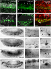
Duf/Kirre, Rols, F-actin, and Blow are expressed in foci during fusion of longitudinal FCs with FCMs. (A–A) Embryo expressing rp298-lacZ in somatic and visceral FCs. (A) Mesodermal cells visualized with anti-β3-Tubulin. Arrowheads point to β-Gal-positive longitudinal FCs. (A) Longitudinal FCs (arrowheads) are mononucleated at this stage. (A) Embryo also expressing sns-NLSmCherry in somatic and visceral FCMs. (B–D) Embryos expressing HLH54F-lacZ. (B) Early stage 12 embryo; longitudinal FCs (arrowheads) still appear to be mononucleated. (C) Embryo in later stage 12, with multinucleated syncytia (arrowheads). (D) Embryo also expressing sns-mCherry; some syncytia appear to be sns-mCherry positive (arrowheads). (E–H) Histochemical staining of wild-type embryos at mid or late stage 13. The embryos are shown as an overview (E, F, G, H) and at two magnifications focusing on longitudinal myogenesis (E, F, G, Hand E, F, G, H). Either heat-fixed embryos (E, F, H) or formaldehyde fixed embryos (G) were stained with anti-Kirre (E–E), anti-Rols7 (F–F), anti-GFP (G–G), or anti-Blow (H–H) antibodies. Arrows point to longitudinal FCs/growing myotubes; arrowheads indicate FCMs. At higher magnifications, local concentrations of the proteins are visible either on the side of the FC/elongation myotube, as in the case of anti-Kirre and anti-Rols7 (E, E; F, F), or in the FCM at the site of attachment for twi::act::GFP(G, G) and anti-Blow (H, H). Scale bars: 20 μm.
Figure 3
Figure 3
Rols7 is transcribed in TVM and circular visceral muscles. (A) Scheme of the rolling pebbles promoter region. rols7: yellow, 3 kb upstream region required for maximum expression in the somatic mesoderm; green, intron with control elements for transcription in the visceral mesoderm and somatic muscles; blue, exons 1 and 2 of rols7. rols6: pink, approximately 1.2 kb upstream region essential for expression in the endoderm and Malpighian tubules; orange, exons 1 and 2. (B) Expression of the roIsIn1-lacZ reporter construct, which contains the regulatory region between exon 1 and 2 of rols7 (green in A). (B’) Magnification of boxed area in (B); β-Gal (green fluorescence) in longitudinal FCs (arrowheads) along the TVM, marked by anti-FasIII (red fluorescence). (C) β-Gal-positive stretching circular muscles (arrow). (D)In situ hybridization of bHLH54F-lacZ embryos using a rols7 probe (green fluorescence). Longitudinal visceral FCs were stained with anti-β-Gal (red fluorescence). (D’ and D”) Magnification of C: arrowheads, rols7 mRNA in β-Gal-negative circular visceral FCs; arrows, rols7 mRNA in β-Gal-positive longitudinal FCs. Scale bars: 20 μm.
Figure 4
Figure 4
Longitudinal muscle fusion requires rols7. Expression of bap-lacZ (green in A, B, E, F) or HLH54F-lacZ (green in GJ; white in C and D) visualized by staining with fluorescent anti-β-Gal and counterstaining with anti-β3-Tubulin (anti-β3Tub red in E, F, H; white in I). Staining of visceral mesoderm with anti-Fasciclin III (anti-FasIII, red in G and J). Lateral view of wild-type embryo (A) and rols mutant embryo (B) at stage 12. Note the β-Gal-positive cells in the rols mutant embryo (B) along the stretching circular muscles. Dorsolateral views of stage 16 wild-type embryo (E) and rols7 mutant embryo (F). (E, F) Arrowheads point to the position of the 1st midgut constriction. (C, D)rols7-deficient embryos stained with anti-β-gal showing (C) mononucleated migrating longitudinal FCs with random protrusions (arrowheads) and (D) morphology of binucleated longitudinal muscles (arrowheads). (G) Anti-FasIII staining of rols7-deficient embryos in mid-embryogenesis; longitudinal FCs located dorsally and ventrally on stretching FasIII-positive circular muscles, which sometimes display small gaps (arrowheads). (H–J) Anterior midgut regions covered with mainly mononucleated longitudinal muscles in different rols alleles at the end of embryogenesis (arrowhead); posterior midgut regions with parallel-orientated longitudinal muscles. Arrows point to regions lacking longitudinal muscles.
Figure 5
Figure 5
Longitudinal muscle development is disturbed in lmd, mbc, blow, and kette mutants. (A–C)lmdE202, (D–F)mbcC1/mbcD112, (G–I)blow2 and blow1/blow2, and (J–L)ketteJ-48/ketteG1–37 mutant embryos carrying the reporter construct HLH54F-lacZ and labeled with anti-β-Gal (green), anti-FasciclinIII (anti-FasIII, red in DF and JL, blue in I) and anti-β3-Tubulin (anti-β3Tub, blue in D, F, JL and red in AC, G, H). (A) Unfused longitudinal muscles in an lmdE202 mutant embryo at stage 14. (B and C) Properly oriented protrusions (arrows in B, inset) and initial midgut chambering (arrow in C) in an lmdE202 mutant embryo at late embryogenesis. (D) Longitudinal visceral muscle migration in a transheterozygous mbcC1/mbcD112 mutant embryo. Arrows point to aberrantly migrating longitudinal FCs. (E–F) Reduction of β-Gal-positive cells and abnormal protrusion formation (arrow in E, inset) in a transheterozygous mbcC1/mbcD112 mutant embryo at stage 16. Arrow in (F) points to region of the midgut not covered by longitudinal FCs. (G) Longitudinal FCs migrating all over the circular muscles in a blow2 mutant embryo during mid-embryogenesis. (H) Mononucleated longitudinal FCs forming protrusions in random directions (double arrow; inset is a magnification of the area) in a blow1/blow2 embryo. (I)blow2 embryo showing defects in constriction formation (arrow) and gaps (arrowheads) between the longitudinal cells; compare to less severe phenotype of blow2/blow1 transheterozygous embryo in (H). (J) Transheterozygous ketteJ4–48/ketteG1–37 mutant embryo with longitudinal FCs along circular muscles. Some cells were not attached to the circular visceral track (arrows). (K and L)ketteJ4–48/ketteG1–37 mutant embryo at the end of embryogenesis, with thin cell protrusions (double arrows in K; inset is a magnification) of the longitudinal FCs. Stretched, mononucleated longitudinal muscles at the end of embryogenesis in blow2(M) and ketteJ4–48(N) mutant embryos. Arrows point to nuclei of longitudinal muscles, marked by anti-DMef2 staining.
Figure 6
Figure 6
scar is required for longitudinal fusion, and FC migration/fusion is promoted by wip. (A–C) Lateral view of stage 16 embryos stained with anti-β3-Tubulin to visualize gut constrictions. (A) Wild-type embryo showing normal gut constrictions (arrow). (B) Homozygous scarΔ37 single and (C)scarΔ37wipf06715 double mutant embryos showing normal gut constrictions, but aberrant gut morphology. (D, F) Homozygous scarΔ37wipf06715 double mutant embryos carrying HLH54F-GFP to mark longitudinal myogenesis. (E) Late stage 15 embryo expressing HLH54F-lacZ in a wild-type background. (D) Stage 13 embryo showing mononucleated myoblasts (arrow) and myoblasts with migration defects (arrowhead). (F) Stage 15 embryo displaying binucleated (two arrowheads) or mononucleated myoblasts (one arrowhead). (G–J) Gene dosage experiments. Embryos were stained with anti-β-Gal, anti-β3-Tubulin, and anti-FasIII. (G, H) Homozygous scarΔ37 mutant embryo carrying HLH54F-GFP and lacking one copy of wipf06715. (G) Late stage 13 embryo with normal longitudinal myoblast migration. Sometimes binucleated cells were seen (arrows). (H) Stage 16 embryo with binucleated gut muscles (arrowheads). (I, J) Homozygous wipf06715 mutant embryo carrying HLH54F-GFP and lacking one copy of scarΔ37. (I) Stage 13 embryo showing aberrant cell migrations (arrowhead) and abnormal protrusion formation (arrow). (J) Stage 15 embryo with binucleated gut muscles (arrows). Scale bars: 50 μm.
Figure 7
Figure 7
Model of myoblast fusion creating the circular and longitudinal visceral muscles and the somatic muscles of the Drosophila embryo. (A) Circular visceral muscles arise by incomplete fusion of one FC (blue nuclei) with one FCM (yellow nuclei, after fusion this nucleus is drawn in brown); this depends on Duf, Rst, Sns, and Mbc. (B) Longitudinal visceral myoblast fusion leads to syncytia, mostly with six nuclei. Duf (blue nuclei) and Sns (yellow nuclei) are specifically expressed according to cell type during this fusion. In the absence of Mbc, no fusion occurs; lack of Rols and Blow leads to a limited number of fusions. (C) During somatic myoblast fusion, lack of Duf, Rst, Sns, and Mbc abolishes fusion almost completely; Rols, Kette, WASp, Wip and Arp3schwächling are required for further fusion events to form individual muscles with their characteristic nuclei number.

References

    1. Abmayr SM, Pavlath GK. Myoblast fusion: lessons from flies and mice. Development. 2012;139:641–656. doi: 10.1242/dev.068353. - DOI - PMC - PubMed
    1. Maqbool T, Jagla K. Genetic control of muscle development: learning from Drosophila. J Muscle Res Cell Motil. 2007;28:397–407. doi: 10.1007/s10974-008-9133-1. - DOI - PubMed
    1. Campos-Ortega JA, Hartenstein V. The embryonic development of Drosophila melanogaster. Berlin: Springer-Verlag; 1985.
    1. Kusch T, Reuter R. Functions for Drosophila brachyenteron and forkhead in mesoderm specification and cell signalling. Development. 1999;126:3991–4003. - PubMed
    1. Goldstein MA, Burdette WJ. Striated visceral muscle of drosophila melanogaster. J Morphol. 1971;134:315–334. doi: 10.1002/jmor.1051340305. - DOI - PubMed

Publication types

MeSH terms

LinkOut - more resources