. 2014 Aug 1;33(15):1667-80.
doi: 10.15252/embj.201387576. Epub 2014 Jul 7.

MicroRNA-125b induces tau hyperphosphorylation and cognitive deficits in Alzheimer's disease

Affiliations

MicroRNA-125b induces tau hyperphosphorylation and cognitive deficits in Alzheimer's disease

Julia Banzhaf-Strathmann et al. EMBO J. .

Abstract

Sporadic Alzheimer's disease (AD) is the most prevalent form of dementia, but no clear disease-initiating mechanism is known. Aβ deposits and neuronal tangles composed of hyperphosphorylated tau are characteristic for AD. Here, we analyze the contribution of microRNA-125b (miR-125b), which is elevated in AD. In primary neurons, overexpression of miR-125b causes tau hyperphosphorylation and an upregulation of p35, cdk5, and p44/42-MAPK signaling. In parallel, the phosphatases DUSP6 and PPP1CA and the anti-apoptotic factor Bcl-W are downregulated as direct targets of miR-125b. Knockdown of these phosphatases induces tau hyperphosphorylation, and overexpression of PPP1CA and Bcl-W prevents miR-125b-induced tau phosphorylation, suggesting that they mediate the effects of miR-125b on tau. Conversely, suppression of miR-125b in neurons by tough decoys reduces tau phosphorylation and kinase expression/activity. Injecting miR-125b into the hippocampus of mice impairs associative learning and is accompanied by downregulation of Bcl-W, DUSP6, and PPP1CA, resulting in increased tau phosphorylation in vivo. Importantly, DUSP6 and PPP1CA are also reduced in AD brains. These data implicate miR-125b in the pathogenesis of AD by promoting pathological tau phosphorylation.

Keywords: Alzheimer's disease; kinases; microRNA‐125b; phosphatases; tau phosphorylation.


Figures

Figure 1
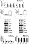
Figure 1. MiR-125b expression alters tau phosphorylation levels in vitro
A Relative miRNA expression was quantified in frontal cortex of five healthy controls and 10 AD cases by TaqMan microRNA assays and normalized to the expression of miR-143 and GAPDH using the ΔΔCt method. N = 5 (controls) or 10 (AD). The box plot depicts the first and third quartile and the median, whiskers: 5–95% percentile, *< 0.05, Mann–Whitney U-test. B Lentiviral miR-125b overexpression in primary hippocampal neurons (DIV12+5) analyzed by TaqMan microRNA assays and normalized to the expression of miR-143 using the ΔΔCt method, n = 3, mean ± SEM, ***< 0.001, Student's t-test. E Transfection of tough decoy (TuD) 125b and a miR-125b sensor construct in primary hippocampal neurons (DIV5+3) analyzed by luciferase assays and normalized to Renilla luciferase expression, n = 6, mean ± SEM, ***< 0.001, Student's t-test. C-G Immunoblots with indicated antibodies (two representative replicates) (C, F) and quantification (D, G) after normalization to total tau or β-actin levels (for total tau). OE: n = 6, TuD: n = 7, mean ± SEM, *< 0.05, **< 0.01, Student's t-test. OE, overexpression; TuD, tough decoy.
Figure 2
Figure 2. MiR-125b regulates tau kinases
Primary hippocampal neurons (DIV12+14) were infected with lentivirus expressing miR-125b or miR-143 as control or tough decoys (TuD) against miR-125b or miR-143. A–D Immunoblots with indicated antibodies (two representative replicates) (A, C) and quantification (B, D) after normalization to either the respective total protein levels (p-p44/42, p-GSK-3β) or β-actin (p35, cdk5, p44/42, GSK-3β). OE: n = 8, TuD: n = 6, mean ± SEM, *< 0.05, Student's t-test. OE, overexpression; TuD, tough decoy.
Figure 3
Figure 3. MiR-125b regulates tau phosphatases
A, B Relative luciferase activity of Bcl-W, DUSP6, PPP1CA, and PPP2CA reporter constructs co-transfected with (A) microRNA-expressing vectors and (B) TuD expressing constructs in primary cortical neurons (DIV5+3). N = 3 with 6 replicates each, mean ± SEM, ***< 0.001, one-way ANOVA. C, D Immunoblots with indicated antibodies (two representative replicates) and quantification after normalization to β-actin levels, n = 6, mean ± SEM, *< 0.05, ***< 0.001, Student's t-test. OE, overexpression; TuD, tough decoy.
Figure 4
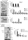
Figure 4. Knockdown of Bcl-W, DUSP6, and PPP1CA phenocopies miR-125b overexpression. Primary hippocampal neurons (DIV12+7) were infected with lentivirus to knock down Bcl-W, DUSP6, PPP1CA (shBcl-W, shDUSP6, shPPP1CA), or a non-targeting control (shControl)
A, B Immunoblots with indicated antibodies (two representative replicates) (A) and quantification of Bcl-W, DUSP6, and PPP1CA (B) after normalization to β-actin levels. N = 6, mean ± SEM, *< 0.05, Student's t-test. C, D Immunoblots with indicated antibodies (two representative replicates) (C) and quantification of phospho-tau (D) after normalization to total tau levels. Total tau levels were normalized to β-actin. N = 4, mean ± SEM, *< 0.05, ***< 0.001, one-way ANOVA. E, F Immunoblots with indicated antibodies (two representative replicates) (E) and quantification of kinase expression (F) after normalization to β-actin or p44/42 (for p-p44/42) levels, n = 4, mean ± SEM, **< 0.01, one-way ANOVA. G Co-expression of miR-125b and HA-Bcl-W/HA-PPP1CA, lacking miR-125b target sites, rescues tau phosphorylation phenotype. Immunoblots with indicated antibodies (two representative replicates). H Quantification of phospho-tau levels from (G) after normalization to total tau, which is normalized to β-actin. N = 6, mean ± SEM, *< 0.05, **< 0.01, ***< 0.001, one-way ANOVA.
Figure 5
Figure 5. Novel miR-125b targets DUSP6 and PPP1CA are reduced in AD brains while kinase levels are elevated
Whole protein lysates from frontal cortex tissue of healthy controls and aged-matched AD patients were analyzed. A, B Representative immunoblots with indicated antibodies. * indicates cross-reactivity with GSK-3α. C Quantification of immunoblots from (A, B) after normalization to either unphosphorylated protein (for AT180, p-p44/42, p-GSK-3β, p-p38, p-SAPK/JNK) or β-actin (for DUSP6, PPP1CA, p35, cdk5, p44/42, GSK-3β, p38, SAPK/JNK). N = 5 (controls), n = 10 (AD), mean ± SEM, *< 0.05, **< 0.01, Mann–Whitney U-test.
Figure 6
Figure 6. High levels of miR-125b impair learning and memory
A Experimental design for administration of miR-125b mimic and the respective negative control (mock) into the dentate gyrus (DG) of wild-type C57BL/6J mice (black arrows) and subsequent fear conditioning (FC) and testing. B C57Bl/6J mice were subjected to contextual fear conditioning, and freezing behavior was analyzed 24 h later. Freezing levels were significantly reduced in miR-125b-injected mice (left panel). The average (avg.) motion index was unchanged (right panel). n = 15–17. C Relative miR-125b expression was quantified in DG, hippocampal CA1 region, and cortex (Ctx) of injected mice by TaqMan microRNA assays and normalization to the expression of miR-143 using the ΔΔCt method. D–F Immunoblots of DG lysates with indicated antibodies (D, E) and quantification (F) after normalization to either unphosphorylated protein (for AT180, p-p44/42, p-GSK-3β, p-p38, p-SAPK/JNK) or β-actin (for Bcl-W, DUSP6, PPP1CA, p35, cdk5, p44/42, GSK-3β, p38, SAPK/JNK). N = 6–8, mean ± SEM, *< 0.05, **< 0.01, Mann–Whitney U-test.

References

    1. Abe M, Bonini N. MicroRNAs and neurodegeneration: role and impact. Trends Cell Biol. 2013;23:30–36. - PMC - PubMed
    1. Absalon S, Kochanek DM, Raghavan V, Krichevsky AM. MiR-26b, upregulated in Alzheimer's disease, activates cell cycle entry, tau-phosphorylation, and apoptosis in postmitotic neurons. J Neurosci. 2013;33:14645–14659. - PMC - PubMed
    1. Alexandrov PN, Dua P, Hill JM, Bhattacharjee S, Zhao Y, Lukiw WJ. microRNA (miRNA) speciation in Alzheimer's disease (AD) cerebrospinal fluid (CSF) and extracellular fluid (ECF) Int J Biochem Mol Biol. 2012;3:365–373. - PMC - PubMed
    1. Andersen CL, Jensen JL, Orntoft TF. Normalization of real-time quantitative reverse transcription-PCR data: a model-based variance estimation approach to identify genes suited for normalization, applied to bladder and colon cancer data sets. Cancer Res. 2004;64:5245–5250. - PubMed
    1. Ballatore C, Lee VM, Trojanowski JQ. Tau-mediated neurodegeneration in Alzheimer's disease and related disorders. Nat Rev Neurosci. 2007;8:663–672. - PubMed

Publication types

MeSH terms