

Quantitative assessment of rest and acetazolamide CBF using quantitative SPECT reconstruction and sequential administration of (123)I-iodoamphetamine: comparison among data acquired at three institutions
- PMID: 25001261
- PMCID: PMC4244544
- DOI: 10.1007/s12149-014-0879-9
Quantitative assessment of rest and acetazolamide CBF using quantitative SPECT reconstruction and sequential administration of (123)I-iodoamphetamine: comparison among data acquired at three institutions
Abstract
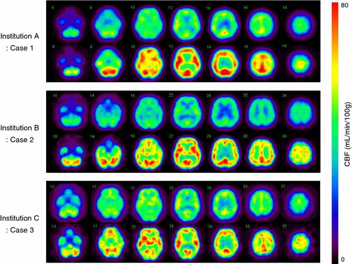
Purpose: A recently developed technique which reconstructs quantitative images from original projection data acquired using existing single-photon emission computed tomography (SPECT) devices enabled quantitative assessment of cerebral blood flow (CBF) at rest and after acetazolamide challenge. This study was intended to generate a normal database and to investigate its inter-institutional consistency.
Methods: The three institutions carried out a series of SPECT scanning on 32 healthy volunteers, following a recently proposed method that involved dual administration of (123)I-iodoamphetamine during a single SPECT scan. Intra-institute and inter-institutional variations of regional CBF values were evaluated both at rest and after acetazolamide challenge. Functional images were pooled for both rest and acetazolamide CBF, and inter-institutional difference was evaluated among these images using two independent software programs.
Results: Quantitative assessment of CBF images at rest and after acetazolamide was successfully achieved with the given protocol in all institutions. Intra-institutional variation of CBF values at rest and after acetazolamide was consistent with previously reported values. Quantitative CBF values showed no significant difference among institutions in all regions, except for a posterior cerebral artery region after acetazolamide challenge in one institution which employed SPECT device with lowest spatial resolution. Pooled CBF images at rest and after acetazolamide generated using two software programs showed no institutional differences after equalization of the spatial resolution.
Conclusions: SPECT can provide reproducible images from projection data acquired using different SPECT devices. A common database acquired at different institutions may be shared among institutions, if images are reconstructed using a quantitative reconstruction program, and acquired by following a standardized protocol.
Figures


References
-
- Minoshima S, Frey KA, Koeppe RA, Foster NL, Kuhl DE. A diagnostic approach in Alzheimer’s disease using three-dimensional stereotactic surface projections of fluorine-18-FDG PET. J Nucl Med. 1995;36(7):1238–1248. - PubMed
-
- Mizumura S, Kumita S, Cho K, Ishihara M, Nakajo H, Toba M, et al. Development of quantitative analysis method for stereotactic brain image: assessment of reduced accumulation in extent and severity using anatomical segmentation. Ann Nucl Med. 2003;17(4):289–295. doi: 10.1007/BF02988523. - DOI - PubMed
Publication types
MeSH terms
Substances
LinkOut - more resources
Full Text Sources
Other Literature Sources
