. 2014 Sep;13(9):1181-90.
doi: 10.1128/EC.00112-14. Epub 2014 Jul 7.

Identification of two nickel ion-induced genes, NCI16 and PcGST1, in Paramecium caudatum

Affiliations

Identification of two nickel ion-induced genes, NCI16 and PcGST1, in Paramecium caudatum

Yasuhiro Takenaka et al. Eukaryot Cell. 2014 Sep.

Abstract

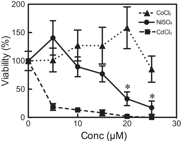
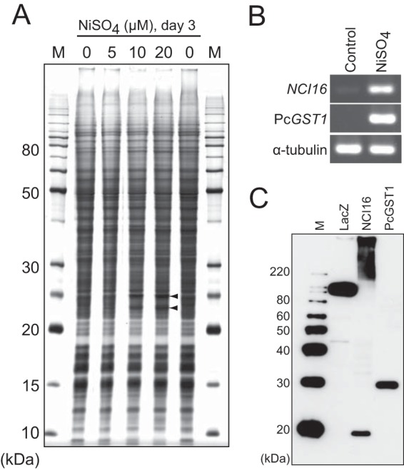
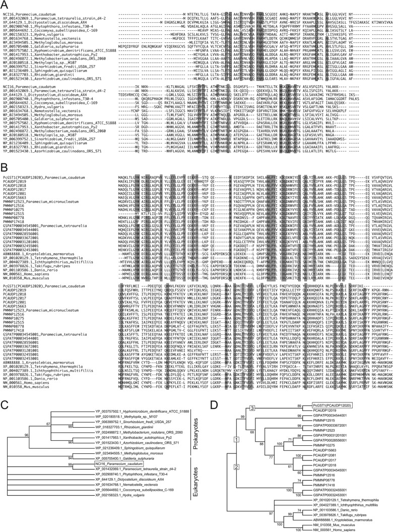
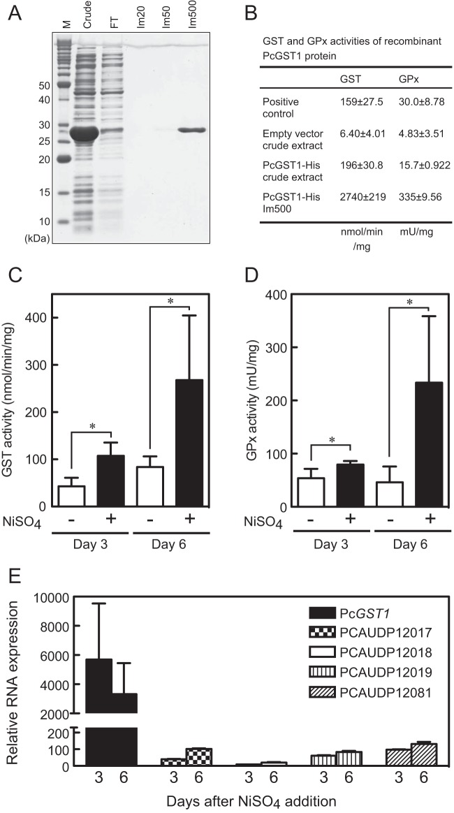
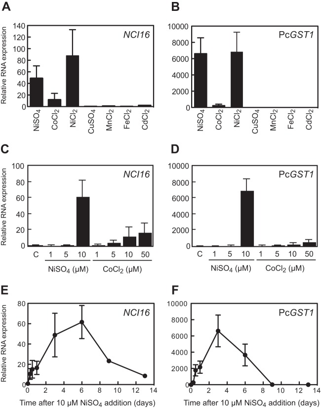
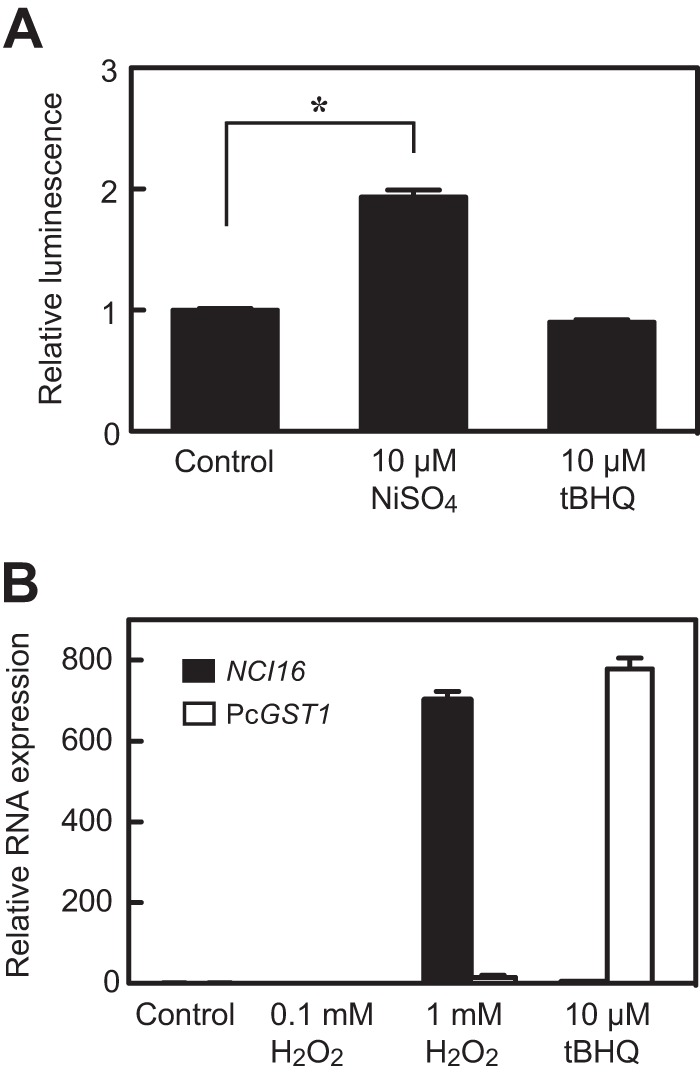
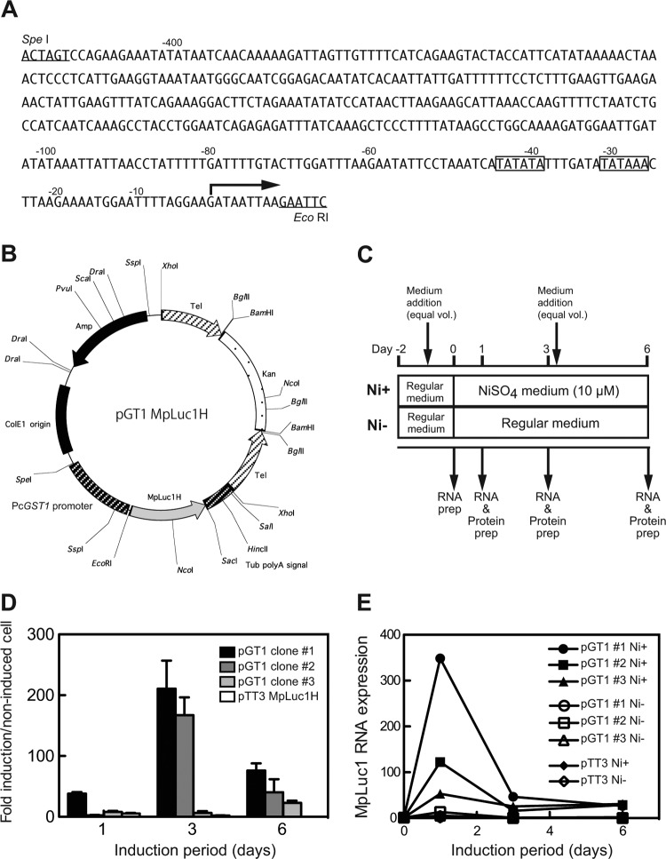
Here, we describe the isolation of two nickel-induced genes in Paramecium caudatum, NCI16 and PcGST1, by subtractive hybridization. NCI16 encoded a predicted four-transmembrane domain protein (∼16 kDa) of unknown function, and PcGST1 encoded glutathione S-transferase (GST; ∼25 kDa) with GST and glutathione peroxidase (GPx) activities. Exposing cells to cobalt chloride also caused the moderate upregulation of NCI16 and PcGST1 mRNAs. Both nickel sulfate and cobalt chloride dose dependently induced NCI16 and PcGST1 mRNAs, but with different profiles. Nickel treatment caused a continuous increase in PcGST1 and NCI16 mRNA levels for up to 3 and 6 days, respectively, and a notable increase in H₂O₂ concentrations in P. caudatum. NCI16 expression was significantly enhanced by incubating cells with H₂O₂, implying that NCI16 induction in the presence of nickel ions is caused by reactive oxygen species (ROS). On the other hand, PcGST1 was highly induced by the antioxidant tert-butylhydroquinone (tBHQ) but not by H2O2, suggesting that different mechanisms mediate the induction of NCI16 and PcGST1. We introduced a luciferase reporter vector with an ∼0.42-kb putative PcGST1 promoter into cells and then exposed the transformants to nickel sulfate. This resulted in significant luciferase upregulation, indicating that the putative PcGST1 promoter contains a nickel-responsive element. Our nickel-inducible system also may be applicable to the efficient expression of proteins that are toxic to host cells or require temporal control.


Figures

FIG 1
FIG 1
Cytotoxicity of NiSO4, CoCl2, and CdCl2 in P. caudatum. Cells were incubated with NiSO4, CoCl2, or CdCl2 at various concentrations (Conc) between 0 and 25 μM for 2 days. Each point indicates the number of treated cells as a percentage ± standard deviations (SD) of untreated cells; moreover, each point represents cell counts from 3 replicate cultures of cells with and without exposure to metal ions. *, P < 0.01 by Student's t test (only for NiSO4).
FIG 2
FIG 2
Identification of nickel ion-induced genes. (A) Identification of induced proteins in whole-cell lysates of P. caudatum incubated with various concentrations of nickel sulfate for 3 days. Cellular proteins (2 μg/lane) were separated by 12.5% SDS-PAGE and silver stained. Arrowheads indicate two major induced proteins. M, BenchMark protein molecular mass marker (Life Technologies). (B) RT-PCR analyses of two nickel-induced genes, NCI16 and PcGST1, after treatment with 10 μM NiSO4 for 3 days. (C) The molecular masses of 10× His-tagged NCI16 and PcGST1 proteins were evaluated by Western blotting probed with anti-His antibody. M, MagicMark XP protein standard (Life Technologies); LacZ, His-tagged LacZ protein as a positive control on Western blotting.
FIG 3
FIG 3
Phylogeny of NCI16 and PcGST1. (A) Multiple-amino-acid sequence alignment of NCI16 and 16 NCI16 orthologs. (B) Multiple-amino-acid sequence alignment of PcGST1 and 21 PcGST1 orthologs. Outlined and shaded text represents at least 90% identical amino acid residues. The ML consensus tree obtained from bootstrap analysis with 1,000 replications of NCI16 (C) and PcGST1 (D) was based on amino acid sequence alignments shown in panels A and B. Bootstrap values of >60% are given to the left of selected nodes. Accession numbers or identifiers used in ParameciumDB are indicated with species names.
FIG 4
FIG 4
GST and GPx activities of PcGST1. (A) Recombinant PcGST1 His-tagged protein was expressed in E. coli, purified by cobalt affinity chromatography, resolved by SDS-PAGE, and stained with CBB. M, protein molecular mass marker. Crude, soluble fraction of E. coli crude extract. FT, flowthrough fraction. Im20, Im50, and Im500, fractions eluted with buffers containing 20, 50, and 500 mM imidazole, respectively. (B) Specific GST and GPx activities of purified recombinant PcGST1. Values for the positive control indicate specific activities of equine liver GST and bovine erythrocyte GPx that are components of GST and GPx assay kits, respectively. Values for the empty vector indicate specific GST and GPx activities of crude extract from E. coli transformed with empty expression vector as a negative control. Specific GST (C) and GPx (D) activities of P. caudatum whole-cell lysate after treatment with 10 μM NiSO4 for 3 or 6 days. Bars show means ± SD from 5 different cultures (*, P < 0.01 by Student's t test). (E) Relative mRNA levels of PcGST1 and 4 other homologs normalized to α-tubulin expression upon nickel exposure. Cells were incubated with 10 μM NiSO4 for 3 or 6 days. Bars represent mean fold changes ± SD (n = 3 per group). The level of mRNA in untreated control cells is defined as 1.0.
FIG 5
FIG 5
Quantitative RT-PCR analyses of NCI16 or PcGST1 mRNA expression. Cells were treated with 10 μM either of the metal ions (1 μM for CdCl2 and CuSO4) for 3 days. Expression of NCI16 (A) or PcGST1 (B) was analyzed by qRT-PCR using gene-specific primers. Values obtained for both genes were normalized to those of P. caudatum α-tubulin mRNA. Concentration-dependent induction of NCI16 (C) or PcGST1 (D) mRNA. Cells were grown in medium containing various concentrations of NiSO4 or CoCl2, harvested 3 days later, and analyzed by qRT-PCR. Induction kinetic of NCI16 (E) or PcGST1 (F) mRNA after treatment with 10 μM NiSO4 also are shown. Bars and points show mean fold changes ± SD from replicate qRT-PCR assays (n = 3) of total RNA from 3 to 4 cell cultures compared to untreated control cells.
FIG 6
FIG 6
Nickel induces ROS production in P. caudatum. (A) Cells were exposed to 10 μM NiSO4 or 10 μM antioxidant tert-butylhydroquinone (tBHQ) for 24 h. Luminescent signals were obtained using an ROS-Glo H2O2 assay kit to measure H2O2 levels. Bars show mean fold changes ± SD from 4 replicate wells compared to 4 untreated control samples (*, P < 0.01, Student's t test). (B) Induction of NCI16 or PcGST1 mRNA in P. caudatum treated with either 1 mM H2O2 or 10 μM tBHQ for 24 h. Bars show mean fold changes ± SD from triplicate qRT-PCR assays compared to untreated control cells.
FIG 7
FIG 7
Analysis of PcGST1 gene promoter. (A) Putative promoter sequence of the PcGST1 gene. Canonical TATA binding boxes are outlined. The bent arrow indicates the putative transcription initiation point. (B) Map of expression vector pGT1-MpLuc1H, carrying the planktonic luciferase gene (MpLuc1) downstream of the SpeI-EcoRI fragment of the PcGST1 gene promoter. Telomere sequences were designed to stabilize a linearized vector in the macronucleus of P. caudatum. (C) Schema of pGT1-MpLuc1H transformant clones incubated with (Ni+) or without (Ni) 10 μM NiSO4. Control (Ni) cells were cultured in regular medium at the same time as Ni+ cells. prep, preparation. (D) Relative luciferase activities were measured in pGT1-MpLuc1H transformant clones at 1, 3, and 6 days after treatment with or without 10 μM NiSO4. Values represent mean fold induction determined from triplicate measurements. The activity of untreated cells is defined as 1.0. (E) Induction kinetics of MpLuc1 mRNA in 3 transformant clones after treatment with (Ni+) or without (Ni) 10 μM NiSO4. Values obtained for MpLuc1 mRNA were normalized to those for P. caudatum α-tubulin mRNA.

References

    1. Scansetti G, Maina G, Botta GC, Bambace P, Spinelli P. 1998. Exposure to cobalt and nickel in the hard-metal production industry. Int. Arch. Occup. Environ. Health 71:60–63. 10.1007/s004200050251 - DOI - PubMed
    1. Sunderman FW., Jr 1989. Mechanisms of nickel carcinogenesis. Scand. J. Work Environ. Health 15:1–12 - PubMed
    1. Denkhaus E, Salnikow K. 2002. Nickel essentiality, toxicity, and carcinogenicity. Crit. Rev. Oncol. Hematol. 42:35–56. 10.1016/S1040-8428(01)00214-1 - DOI - PubMed
    1. Athar M, Hasan SK, Srivastava RC. 1987. Evidence for the involvement of hydroxyl radicals in nickel mediated enhancement of lipid peroxidation: implications for nickel carcinogenesis. Biochem. Biophys. Res. Commun. 147:1276–1281. 10.1016/S0006-291X(87)80208-5 - DOI - PubMed
    1. Cavallo D, Ursini CL, Setini A, Chianese C, Piegari P, Perniconi B, Iavicoli S. 2003. Evaluation of oxidative damage and inhibition of DNA repair in an in vitro study of nickel exposure. Toxicol. In Vitro 17:603–607. 10.1016/S0887-2333(03)00138-3 - DOI - PubMed

MeSH terms

LinkOut - more resources