. 2014 Nov 15;210(10):1658-69.
doi: 10.1093/infdis/jiu321. Epub 2014 Jun 13.

An ahemolytic pneumolysin of Streptococcus pneumoniae manipulates human innate and CD4⁺ T-cell responses and reduces resistance to colonization in mice in a serotype-independent manner

Affiliations

An ahemolytic pneumolysin of Streptococcus pneumoniae manipulates human innate and CD4⁺ T-cell responses and reduces resistance to colonization in mice in a serotype-independent manner

M Nadeem Khan et al. J Infect Dis. .

Abstract

Background: Some Streptococcus pneumoniae serotypes express an ahemolytic pneumolysin (PLYa). Serotypes that commonly express PLYa, including serotype 8 (ST8) and ST1, are often associated with a low prevalence during colonization but a higher propensity to cause invasive disease. We sought to study the host response to ST8 PLYa in a homologous and heterologous capsular background.

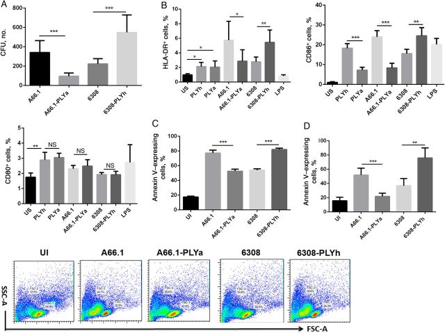
Methods: We genetically exchanged the PLYa of ST8 strain 6308 with the hemolytic PLY (PLYh) of ST3 A66.1 and vice versa and determined the impact of the exchange on nasopharyngeal colonization in mice. Then, to compare the response of human cells to PLYa-expressing and PLYh-expressing strains, we infected human peripheral blood mononuclear cells (PBMCs) with PLY-switched strains and assessed dendritic cell and CD4(+) T-cell responses by intracellular cytokine staining.

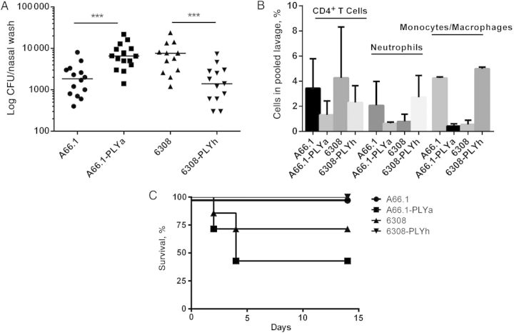
Result: Mice colonized with PLYa-expressing strains had significantly higher colonization densities than those colonized with PLYh-expressing strains, irrespective of capsular background. Compared with infection of PBMCs with PLYh-expressing strains, infection with PLYa-expressing strains induced diminished innate (dendritic cell cytokines, costimulatory receptor, and apoptotic) and adaptive (CD4(+) T-cell proliferative and memory interleukin 17A) responses.

Conclusion: Our findings demonstrate that PLYa has the potential to manipulate host immunity irrespective of capsule type. PLY exchange between STs expressing PLYa and PLYh could lead to unexpected colonization or invasion phenotypes.

Keywords: Streptococcus pneumoniae; T cell; apoptosis; colonization; dendritic cell; invasion; mice; pneumolysin; serotype.


Figures

Figure 1.
Figure 1.
Construction and characterization of pneumolysin (PLY)–switched serotype 3 (ST3) and ST8 strains. A, Design of gene construct used for the construction of PLY-switched strains. B, Confirmation of PLY-switched ST3 and ST8 strains by hemolytic assay. Data represent mean (+SD) of 1 experiment in duplicate. C, Western blot of in vitro–grown PLY-switched strains. Lane 1, Hemolytic PLY (PLYh); lane 2, A66.1; lane 3, A66.1–ahemolytic PLY (PLYa); lane 4, 6308; lane 5, 6308-PLYh; lane 6, 6308-PLYa; lane 7, PLY knockout. D, Quantitation of ST3 and ST8 polysaccharides in in vitro–grown culture supernatants and pellets by enzyme-linked immunosorbent assay. Data is representative of 1 experiment in triplicate and expressed as mean (+SD). Abbreviation: NS, not significant.
Figure 2.
Figure 2.
Nasopharyngeal (NP) colonization of mice with pneumolysin (PLY)–switched serotype 3 (ST3) and ST8 strains. A, Five mice per group were intranasally infected with ST3 A66.1 or A66.1–ahemolytic PLY (PLYa; 105 colony-forming units [CFU]/mouse) or ST8 6308, 6308–hemolytic PLY (PLYh), and 6308-PLYa (5 × 10⁴ CFU each). The number of CFU (in nasopharyngeal lavage) on day 7 after colonization is shown. Data were analyzed by analysis of variance (P < .001,) followed by the Dunn post hoc test for differences between wild-type and PLY-switched strains. Data are representative of 3 separate experiments (5 mice each) and represented as medians. ***P < .001. B, CD4+ T cells, neutrophils, and monocytes/macrophages in the pooled nasal lavage of mice colonized with PLY-switched ST3 and ST8 strains. Cells were gated on lymphocytes based on forward and side scatter, followed by the exclusion of doublets. The CD3 population was gated for Ly6G+Cd11b+ cells and considered to be neutrophils. CD11bF480+ cells were considered to be macrophages/monocytes, and CD3+CD4+ double-positive cells were considered to be CD4+ T cells. Data represent mean (+SD) of 3 experiments (each in singlet). For differences in each cell type (CD4+ T cells, neutrophils, or monocytes/macrophages) between wild-type and respective PLY-switched group (ST3 or ST8), data were analyzed by the Student t test. ***P < .001. C, In a separate experiment, 7 mice per group were infected with a higher inoculum (5 × 10⁶ CFU) of wild-type or PLY-switched ST3 and ST8 strains. Data were analyzed by the Mantel–Cox test. *P = .022.
Figure 3.
Figure 3.
Cytokine response in human myeloid dendritic cells (mDCs) by intracellular cytokine staining (ICS). Peripheral blood mononuclear cells (PBMCs) were rested overnight and then either stimulated with hemolytic pneumolysin (PLYh) or ahemolytic PLY (PLYa; 500 ng/mL) or infected with PLY-switched serotype 3 (ST3) and ST8 strains at a multiplicity of infection of 1:10 for 2 hours. After 2 hours, extracellular bacteria were killed, brefeldin A was added to the culture, and cells were further incubated for an additional 5 hours (total incubation time, 7 hours). Cytokine response was detected in HLA-DR+ CD11chigh DCs by ICS. A, Initial gating of lymphocytes, based on forward-scatter and side-scatter properties. CD3, DAPI, and CD66b were used as a single-color (Pacific blue) dump channel to exclude dead cells, T cells, and granulocytes, followed by the exclusion of cell doublets, using forward-scatter area (FSC-A) versus forward-scatter height (FSC-H) parameters. The CD3CD66bDAPI population was further gated for CD14+HLA-DR+ to identify monocytes. CD14 and HLA-DR+ cells were subsequently gated for CD11c+ and enumerated as mDCs (HLA-DR+CD11chigh). Gates for cytokine-positive DCs were then determined, based on observations with unstimulated cells (PBMCs plus medium) and stimulated cells (PBMCs plus pneumococcal strains or lipopolysaccharide). Cells were acquired on LSR II (BD). B, Cytokine response in mDCs. Data are representative of 5 separate experiments, and each experiment was performed in triplicate (n = 5 donors). Data were analyzed by analysis of variance (ANOVA) and the Dunn post hoc test. **P < .01 and ***P < .001. C, Toll-like receptor 2 (TLR-2) and TLR-4 blocking and cytokine response in DCs. Anti–TLR-2 and anti–TLR-4 monoclonal antibodies were used to neutralize TLR-2 and TLR-4 in PBMCs, which were subsequently stimulated with either PLYh or PLYa. Data are representative of 3 separate experiments, and each experiment was performed in triplicate (n = 3 donors). Data were analyzed by ANOVA and the Dunn post hoc test. *P < .05, **P < .01, and ***P < .001. Abbreviation: NS, not significant.
Figure 4.
Figure 4.
Uptake of pneumolysin (PLY)–switched serotype 3 (ST3) and ST8 strains by dendritic cells (DCs) and impact on maturation and apoptosis. DCs were derived in vitro from magnetically sorted CD14+ monocytes in the presence of granulocyte-macrophage colony-stimulating factor and interleukin 4 and subsequently stimulated with hemolytic PLY or ahemolytic PLY or infected with PLY-switched ST3 and ST8 strains at a multiplicity of infection of 1:10. Data were analyzed by analysis of variance and the Dunn post hoc test. *P < .05, **P < .01, and ***P < .001. A, Uptake of PLY-switched ST3 and ST8 strains by DCs. Data are representative of 3 separate experiments in triplicate (n = 3 donors) and represented as mean + SD. B, DCs were infected with PLY-switched ST3 and ST8 strains, and expression of maturation markers was studied on DCs. Data are representative of 3 separate experiments in triplicate (n = 3 donors) and are represented as mean + SD. C, DCs were infected with PLY-switched ST3 and ST8 strains, and apoptosis was studied by staining cells with annexin V antibody. Data are representative of 3 separate experiments in triplicate (n = 3 donors) and are represented as mean + SD. D, Peripheral blood mononuclear cells (PBMCs) were infected with PLY-switched ST3 and ST8 strains, and apoptosis was studied by staining cells with annexin V antibody. Data are representative of 3 separate experiments in triplicate (n = 3 donors) and are represented as mean + SD. E, A comparison of forward scatter (FSC) shrinkage in PBMCs infected with PLY-switched ST3 and ST8 strains. Abbreviations: CFU, colony-forming unit; NS, not significant; SSC, side scatter.
Figure 5.
Figure 5.
CD4+ T-cell proliferation. CFSE-stained splenocytes (1 million per stimulation) were stimulated with hemolytic pneumolysin (PLYh), ahemolytic PLY (PLYa), or PLY-switched serotype 3 (ST3) and ST8 strains for 10 days. Stimulated cells were stained with anti-CD4 antibody (APC), and cells were acquired on flow cytometry (LSR II; BD). A, Representative dot plots from cells stimulated with PLY-switched ST3 and ST8 strains. B, CD4+ T-cell proliferation percentage. Data are mean + SD of 5 separate experiments (n = 5 donors). Each experiment was performed in triplicate. Data were analyzed by analysis of variance, and the Dunn post hoc test. *P < .05.
Figure 6.
Figure 6.
Frequency of memory interleukin 17A (IL-17A)–expressing CD4+ T-cell response. Peripheral blood mononuclear cells (PBMCs) were rested overnight and then stimulated with hemolytic pneumolysin (PLYh) or ahemolytic PLY (PLYa; 500 ng/mL each) or infected with PLY-switched serotype 3 (ST3) and ST8 strains at a multiplicity of infection of 1:10 for 2 hours. After 2 hours, extracellular bacteria were killed, brefeldin A was added to the culture, and cells were further incubated for an additional 5 hours (total incubation time, 7 hours). The IL-17A response was detected in CD4+ T cells by intracellular cytokine staining. A, Cells were gated on lymphocytes on the basis of forward scatter (FSC) and side scatter (SSC), followed by the exclusion of doublets and dead cells (7-AAD+). Live cells were further gated, with CD3+CD4+ double-positive T cells representing the CD4+ T cells. IL-17A–expressing CD4+ T cells were further gated against CD45 RA memory marker. The percentage of responding cells was calculated by dividing frequencies of cytokine-positive cells by total CD4+ T-cell counts for the respective stimulation. Representative dot plots from cells stimulated with PLY-switched ST3 and ST8 strains. B, Frequency of IL-17A–expressing memory CD4+ T cells (mean + SD) of 5 separate experiments (n = 5 donors). Each experiment was performed in triplicate. Data were analyzed by analysis of variance, and the Dunn post hoc test. *P < .05 and **P < .01.

Similar articles

Cited by

References

    1. Ogunniyi AD, LeMessurier KS, Graham RM, et al. Contributions of pneumolysin, pneumococcal surface protein A (PspA), and PspC to pathogenicity of Streptococcus pneumoniae D39 in a mouse model. Infect Immun. 2007;75:1843–51. - PMC - PubMed
    1. Richards L, Ferreira DM, Miyaji EN, Andrew PW, Kadioglu A. The immunising effect of pneumococcal nasopharyngeal colonisation; protection against future colonisation and fatal invasive disease. Immunobiology. 2010;215:251–63. - PubMed
    1. Malley R, Henneke P, Morse SC, et al. Recognition of pneumolysin by Toll-like receptor 4 confers resistance to pneumococcal infection. Proc Natl Acad Sci U S A. 2003;100:1966–71. - PMC - PubMed
    1. Witzenrath M, Pache F, Lorenz D, et al. The NLRP3 inflammasome is differentially activated by pneumolysin variants and contributes to host defense in pneumococcal pneumonia. J Immunol. 2011;187:434–40. - PubMed
    1. Melin M, Trzcinski K, Antonio M, et al. Serotype-related variation in susceptibility to complement deposition and opsonophagocytosis among clinical isolates of Streptococcus pneumoniae. Infect Immun. 2010;78:5252–61. - PMC - PubMed

Publication types