. 2014 Aug 28;124(9):1481-91.
doi: 10.1182/blood-2014-05-574830. Epub 2014 Jul 7.

PKC-β as a therapeutic target in CLL: PKC inhibitor AEB071 demonstrates preclinical activity in CLL

Affiliations

PKC-β as a therapeutic target in CLL: PKC inhibitor AEB071 demonstrates preclinical activity in CLL

Dalia El-Gamal et al. Blood. .

Abstract

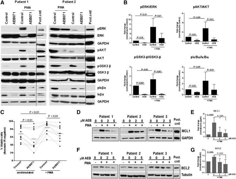
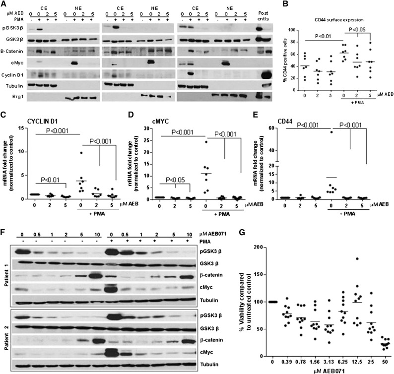
Targeting B-cell receptor (BCR) signaling in chronic lymphocytic leukemia (CLL) has been successful with durable remissions observed with several targeted therapeutics. Protein kinase C-β (PKC-β) is immediately downstream of BCR and has been shown to be essential to CLL cell survival and proliferation in vivo. We therefore evaluated sotrastaurin (AEB071), an orally administered potent PKC inhibitor, on CLL cell survival both in vitro and in vivo. AEB071 shows selective cytotoxicity against B-CLL cells in a dose-dependent manner. Additionally, AEB071 attenuates BCR-mediated survival pathways, inhibits CpG-induced survival and proliferation of CLL cells in vitro, and effectively blocks microenvironment-mediated survival signaling pathways in primary CLL cells. Furthermore, AEB071 alters β-catenin expression, resulting in decreased downstream transcriptional genes as c-Myc, Cyclin D1, and CD44. Lastly, our preliminary in vivo studies indicate beneficial antitumor properties of AEB071 in CLL. Taken together, our results indicate that targeting PKC-β has the potential to disrupt signaling from the microenvironment contributing to CLL cell survival and potentially drug resistance. Future efforts targeting PKC with the PKC inhibitor AEB071 as monotherapy in clinical trials of relapsed and refractory CLL patients are warranted.


Figures

Figure 1
Figure 1
AEB071 induces selective cytotoxicity in CLL cells and inhibits proliferation. (A-B) CD19+ cells from CLL patients (N = 11) were incubated with or without increasing concentrations of AEB071 for up to 72 hours. Viability was determined by MTS assay and was calculated relative to time-matched untreated controls. Dark lines represent averages. (C) Whole blood from CLL patients (N = 5) was incubated with AEB071 (2 μM) for 24 hours. Viability was determined by flow cytometry as described in “Methods.” Dark lines represent averages. (D) Whole blood from normal subjects (N = 5) was incubated without AEB071 (2 μM) for 24 hours. Viability was determined by flow cytometry as described in “Methods.” Dark lines represent averages. (E) CD19+ cells from CLL patients (N = 3) were treated with AEB071 (2 μM) and stimulated with CpG685 oligonucleotides (3.2 μM) for 24 hours. AKT phosphorylation at Ser473, GSK3-β phosphorylation at Ser9, and ERK phosphorylation at Thr202/Tyr204 was assessed by immunoblot. A representative blot with band quantification is presented; black line indicates cropped regions wherein only relevant bands are shown. (F) CD19+ cells from CLL patients (N = 17) were incubated with AEB071 (1 μM) and CpG685 (3.2 μM) for 72 hours. Viability was determined by MTS. Dark lines represent averages. (G) CD19+ cells from CLL patients (N = 10) were incubated with CpG685 (3.2 μM) and treated with AEB071 (1 or 2 μM) or PCI-32765 (1 μM) and proliferation was assessed 120 hours later by tritiated thymidine. Dark lines represent averages.
Figure 2
Figure 2
AEB071 attenuates BCR-dependent activation. (A-B) CD19+ cells from CLL patients (N = 8) were treated with AEB071 (2 μM) and stimulated with immobilized anti-IgM for 24 hours. ERK phosphorylation at Thr202/Tyr204, AKT phosphorylation at Ser473, GSK3-β phosphorylation at Ser9, and IκBα phosphorylation was assessed by immunoblot. Results from 2 patients are presented; black line indicates cropped regions wherein only relevant bands are shown. Band quantification is represented as fold change from unstimulated control (mean ± standard deviation [SD]). (C) CD19+ cells from CLL patients (N = 11) were stimulated with immobilized anti-IgM in presence or absence of AEB071 (2 μM) for 24 hours and cytotoxicity was measured by annexin/PI staining. Dark lines represent averages.
Figure 3
Figure 3
AEB071 antagonizes PMA-induced B-CLL survival and enhances apoptosis. (A-B) CD19+ cells from CLL patients (N = 7) were treated with AEB071 (2 μM) and stimulated with PMA (250 ng/mL) for 24 hours. ERK phosphorylation at Thr202/Tyr204, AKT phosphorylation at Ser473, GSK3-β phosphorylation at Ser9, and IκBα phosphorylation was assessed by immunoblot. Results from 2 patients are presented; black line indicates cropped regions wherein only relevant bands are shown. Band quantification is represented as fold change from unstimulated control (mean ± SD). (C) CD19+ cells from CLL patients (N = 10) were stimulated with PMA (250 ng/mL) in presence or absence of AEB071 (2 μM) for 24 hours and cytotoxicity was measured by annexin/PI staining. Dark lines represent averages. (D-G) CD19+ cells from CLL patients (N = 6) were treated with AEB071 (2 or 5 μM) and stimulated with PMA (250 ng/mL) for 24 hours. Expression of MCL1 (D-E) and BCL2 (F-G) was assessed by immunoblot. Results from 3 patients are presented; black line indicates cropped regions wherein only relevant bands are shown. Band quantification is represented as fold change from unstimulated control (mean ± SD).
Figure 4
Figure 4
AEB071 antagonizes microenvironment stimuli and stromal-induced protection. CD19+ cells from CLL patients (N = 12-21) were incubated with 1 μM AEB071 and 1 μg/mL CD40L (A), 50 ng/mL BAFF (B), 20 ng/mL TNF-α (C), 800 U/mL IL-4 (D), and 100 ng/mL CXCL12 (E). Viability at 72 hours was determined by MTS. Dark lines represent averages. (F) CD19+ cells from CLL patients (N = 7) were isolated from peripheral blood and incubated with AEB071 (1 μM) in suspension or in coculture with 9-15c stromal cells for 48 hours. Viability was determined by annexin V/PI flow cytometry. Dark lines represent averages.
Figure 5
Figure 5
GSK3-β contributes to AEB071-induced effects in B-CLL. (A-B) CD19+ cells from CLL patients (N = 9) were treated with AEB071 (2 μM) and stimulated with immobilized anti-IgM for 30 minutes or 24 hours. PKC phosphorylation at Thr638/641 and GSK3-β phosphorylation at Ser9 was assessed by immunoblot. Results from 2 patients are shown and band quantification is represented as fold change from unstimulated control (mean ± SD). (C-D) CD19+ cells from CLL patients (N = 6) were pretreated with media or okadaic acid (5 nM) for 2 hours, followed by incubation for 3 hours with AEB071 (2 or 5 μM). GSK3-β phosphorylation at Ser9 and AKT phosphorylation at Ser473 was assessed by immunoblot. Results from 2 patients are shown and GSK3-β band quantification is represented as fold change from unstimulated control (mean ± SD). (E) CD19+ cells from CLL patients (N = 4) were pretreated with media or okadaic acid (5 nM) for 2 hours, followed by incubation with AEB071 for 48 hours. Cell viability was measured by annexin/PI staining and expressed as mean ± SD. (F) CD19+ cells from CLL patients (N = 3) were treated with AEB071 (2 μM) or FTY720 for 5 hours. Enzymatic activity of PP2A in the cell lysates was measured by a nonradioactive assay as described in methods and expressed as fold change from untreated control (mean ± SD).
Figure 6
Figure 6
AEB071 targets β-catenin and its downstream transcriptional targets in B-CLL. (A) CD19+ cells from CLL patients (N = 3) were treated with AEB071 (2 or 5 μM) and stimulated with PMA (250 ng/mL) for 24 hours. GSK3-β phosphorylation at Ser9 and protein expression of β-Catenin, c-Myc, and Cyclin D1 was assessed in cytoplasmic (CE) and nuclear extracts (NE) by immunoblot. (B-E) CD19+ cells from CLL patients (N = 7) were treated with AEB071 (2 or 5 μM) and stimulated with PMA (250 ng/mL) for 24 hours, wherein surface expression of CD44 (B) was evaluated in viable cells by flow cytometry and transcript levels of Cyclin D1 (C), c-Myc (D), and CD44 (E) was estimated by real-time PCR. Dark lines represent averages. (F) CD19+ cells from CLL patients (N = 3) were treated with increasing concentrations of AEB071 and stimulated with PMA (250 ng/mL) for 24 hours. GSK3-β phosphorylation at Ser9 and protein expression of β-Catenin and c-Myc was assessed in whole-cell lysates by immunoblot. Results from 2 patients are presented. (G) CD19+ cells from CLL patients (N = 10) were incubated with increasing concentrations of AEB071 for 24 hours. Viability was determined by MTS assay and calculated relative to untreated control. Dark lines represent averages.

References

    1. Woyach JA, Johnson AJ, Byrd JC. The B-cell receptor signaling pathway as a therapeutic target in CLL. Blood. 2012;120(6):1175–1184. - PMC - PubMed
    1. Burger JA, Gribben JG. The microenvironment in chronic lymphocytic leukemia (CLL) and other B cell malignancies: insight into disease biology and new targeted therapies. Semin Cancer Biol. 2014;24:71–81. - PubMed
    1. Zhang S, Kipps TJ. The pathogenesis of chronic lymphocytic leukemia. Annu Rev Pathol. 2014;9:103–118. - PMC - PubMed
    1. Herman SE, Gordon AL, Wagner AJ, et al. Phosphatidylinositol 3-kinase-δ inhibitor CAL-101 shows promising preclinical activity in chronic lymphocytic leukemia by antagonizing intrinsic and extrinsic cellular survival signals. Blood. 2010;116(12):2078–2088. - PMC - PubMed
    1. Herman SE, Lapalombella R, Gordon AL, et al. The role of phosphatidylinositol 3-kinase-δ in the immunomodulatory effects of lenalidomide in chronic lymphocytic leukemia. Blood. 2011;117(16):4323–4327. - PMC - PubMed

Publication types

MeSH terms