. 2014 Jul 7;15(1):572.
doi: 10.1186/1471-2164-15-572.

Optimizing multiplex SNP-based data analysis for genotyping of Mycobacterium tuberculosis isolates

Affiliations

Optimizing multiplex SNP-based data analysis for genotyping of Mycobacterium tuberculosis isolates

Sarah Sengstake et al. BMC Genomics. .

Abstract

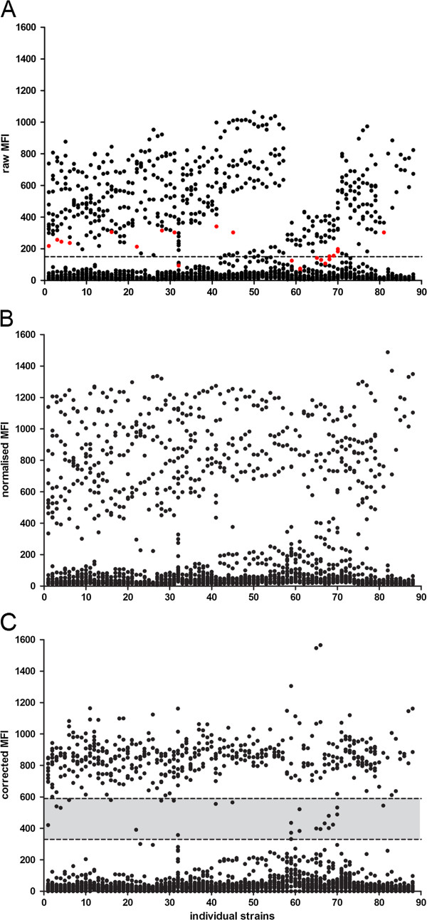
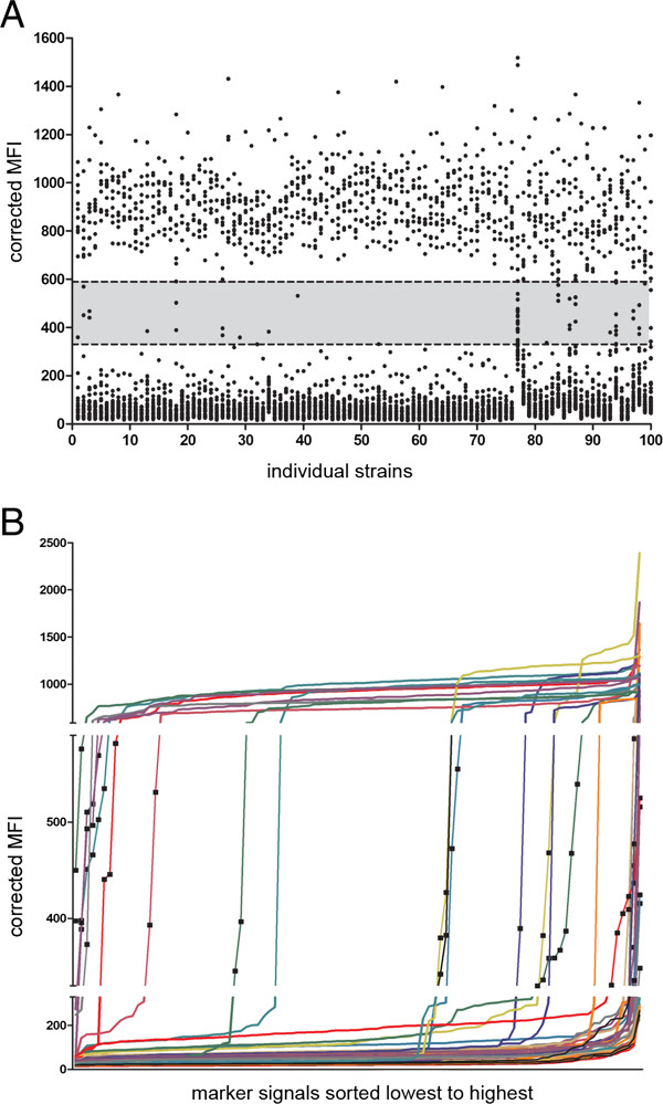
Background: Multiplex ligation-dependent probe amplification (MLPA) is a powerful tool to identify genomic polymorphisms. We have previously developed a single nucleotide polymorphism (SNP) and large sequence polymorphisms (LSP)-based MLPA assay using a read out on a liquid bead array to screen for 47 genetic markers in the Mycobacterium tuberculosis genome. In our assay we obtain information regarding the Mycobacterium tuberculosis lineage and drug resistance simultaneously. Previously we called the presence or absence of a genotypic marker based on a threshold signal level. Here we present a more elaborate data analysis method to standardize and streamline the interpretation of data generated by MLPA. The new data analysis method also identifies intermediate signals in addition to classification of signals as positive and negative. Intermediate calls can be informative with respect to identifying the simultaneous presence of sensitive and resistant alleles or infection with multiple different Mycobacterium tuberculosis strains.

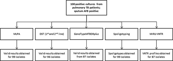
Results: To validate our analysis method 100 DNA isolates of Mycobacterium tuberculosis extracted from cultured patient material collected at the National TB Reference Laboratory of the National Center for Tuberculosis and Lung Diseases in Tbilisi, Republic of Georgia were tested by MLPA. The data generated were interpreted blindly and then compared to results obtained by reference methods. MLPA profiles containing intermediate calls are flagged for expert review whereas the majority of profiles, not containing intermediate calls, were called automatically. No intermediate signals were identified in 74/100 isolates and in the remaining 26 isolates at least one genetic marker produced an intermediate signal.

Conclusion: Based on excellent agreement with the reference methods we conclude that the new data analysis method performed well. The streamlined data processing and standardized data interpretation allows the comparison of the Mycobacterium tuberculosis MLPA results between different experiments. All together this will facilitate the implementation of the MLPA assay in different settings.


Figures

Figure 1
Figure 1
Stepwise approach of the data analysis method. Dot blots illustrate MFI values for 43 genetic markers targeted in 88 clinical isolates and laboratory strains [10] obtained from (A) the MAGPIX csv file, (B) after normalization and (C) after normalization and correction. (A) Raw MFI values obtained for every targeted marker per strain. The dashed line indicates the threshold of MFI 150 which was initially chosen for the classification of targeted makers. Red dots show the MFI values obtained which are located in the intermediate range after normalization and correction in panel C. (B) MFI values after intra-strain normalization of raw MFI values. (C) MFI signals after normalization and inter-strain correction using marker-specific correction factors. The grey area defines the intermediate range calculated as the area between one and two standard deviations from the average MFINORM = 860.
Figure 2
Figure 2
Visualization of data generated from 100 Georgian isolates after data normalization and data correction. (A) Dot plot showing normalized and corrected MFI values (black dots) per isolate for all 4300 markers targeted in the 100 Georgian isolates. The grey area highlights 62 (1.4%) unclassifiable markers of which 24 are drug resistance markers. Markers located above this area are classified as positive (971, 22.6%) and below as negative (3267, 76%). (B) Same data as shown in (A) but only the intermediate values are shown and visualized per marker. Each line shows the distribution of normalized and corrected MFI values, sorted from lowest o highest, (black squares), for one marker (individual colors).
Figure 3
Figure 3
Results obtained of the 100 isolates by various methods. Samples were taken from 100 individual patients, all diagnosed with pulmonary TB and producing AFB positive sputum smear; MLPA, DST, GenoTypeMTBDRplus, spoligotyping and MIRU-VNTR was performed on all 100 isolates. DST for first line drugs was performed on all 100 isolates whereas DST for second line drugs was performed on drug-resistant TB isolates only. DST results were not available for four isolates due to contamination of the respective cultures; No information was obtained from three isolates tested by GenoTypeMTBDRplus; Spoligotypes were not obtained for one isolate. Unknown spoligotypes were obtained for 17 isolates and the spoligotypes of five isolates were not reported in the SITVITWEB database. MIRU-VNTR types were not obtained for 13 isolates. Multiple copy numbers in one or more loci were revealed in two isolates; Genotypic information of 45 markers screened per isolate was obtained for all 100 isolates by MLPA. For 85 isolates, lineage types could be assigned on the basis of consistent lineage marker profiles and in 14 isolates after expert review. For one isolate the lineage type profile was not interpretable. For 46 isolates molecular drug resistance was identified by MLPA of which 11 isolates had intermediate signals for at least one drug resistance conferring marker.

Similar articles

Cited by

References

    1. Borgdorff MW, van Soolingen D. The re-emergence of tuberculosis: what have we learnt from molecular epidemiology? Clin Microbiol Infect. 2013;19:889–901. doi: 10.1111/1469-0691.12253. - DOI - PubMed
    1. Devaux I, Kremer K, Heersma H, van Soolingen D. Clusters of multidrug-resistant Mycobacterium tuberculosis cases, Europe. Emerg Infect Dis. 2009;15:1052–1060. doi: 10.3201/eid1507.080994. - DOI - PMC - PubMed
    1. Dalla Costa ER, Lazzarini LC, Perizzolo PF, Diaz CA, Spies FS, Costa LL, Ribeiro AW, Barroco C, Schuh SJ, da Silva Pereira MA, Dias CF, Gomes HM, Unis G, Zaha A, da Silva PE A, Suffys PN, Rossetti ML. Mycobacterium tuberculosis of the RDRio genotype is the predominant cause of tuberculosis and associated with multidrug resistance in Porto Alegre City South Brazil. J Clin Microbiol. 2013;51:1071–1077. doi: 10.1128/JCM.01511-12. - DOI - PMC - PubMed
    1. Monteserin J, Camacho M, Barrera L, Palomino JC, Ritacco V, Martin A. Genotypes of Mycobacterium tuberculosis in patients at risk of drug resistance in Bolivia. Infect Genet Evol. 2013;17:195–201. doi: 10.1016/j.meegid.2013.04.010. - DOI - PubMed
    1. Gagneux S, DeRiemer K, Van T, Kato-Maeda M, de Jong BC, Narayanan S, Nicol M, Niemann S, Kremer K, Gutierrez MC, Hilty M, Hopewell PC, Small PM. Variable host-pathogen compatibility in Mycobacterium tuberculosis. Proc Natl Acad Sci U S A. 2006;103:2869–2873. doi: 10.1073/pnas.0511240103. - DOI - PMC - PubMed

Publication types

MeSH terms