. 2014 Sep;82(9):3927-38.
doi: 10.1128/IAI.01779-14. Epub 2014 Jul 7.

Brucella melitensis invades murine erythrocytes during infection

Affiliations

Brucella melitensis invades murine erythrocytes during infection

Marie-Alice Vitry et al. Infect Immun. 2014 Sep.

Abstract

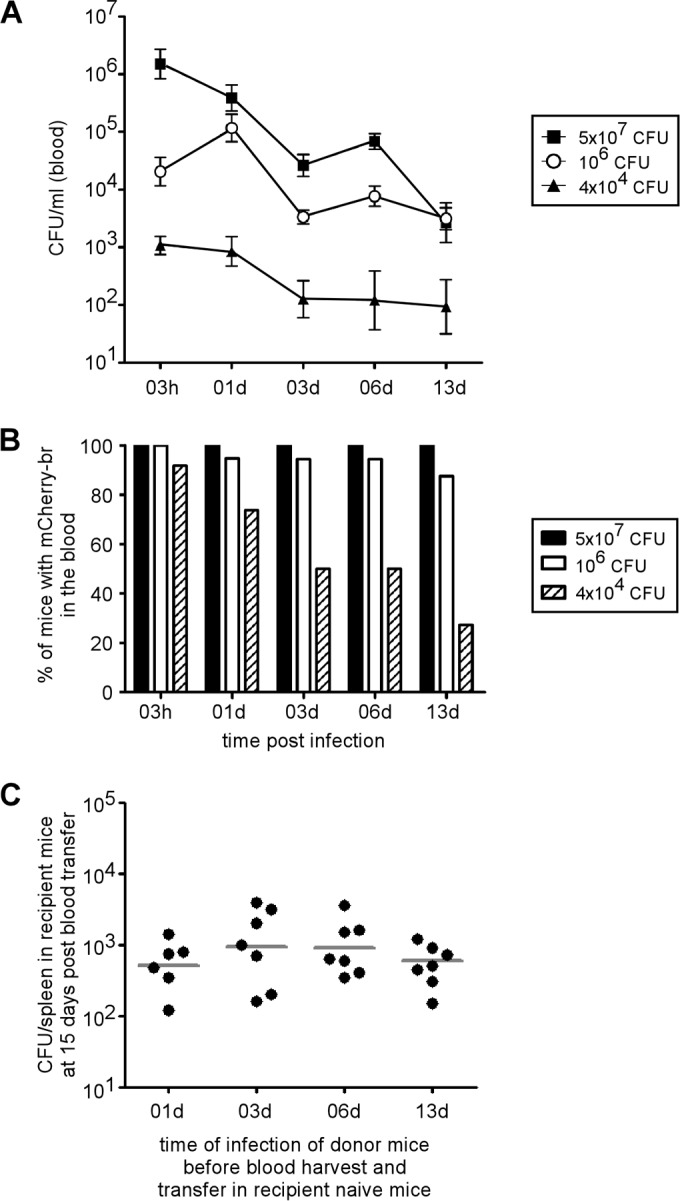
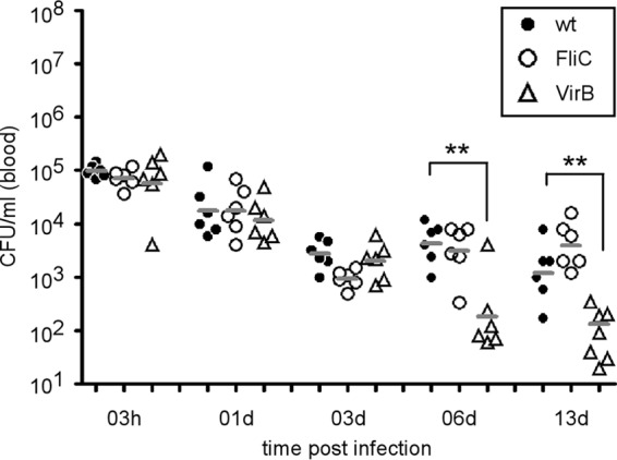
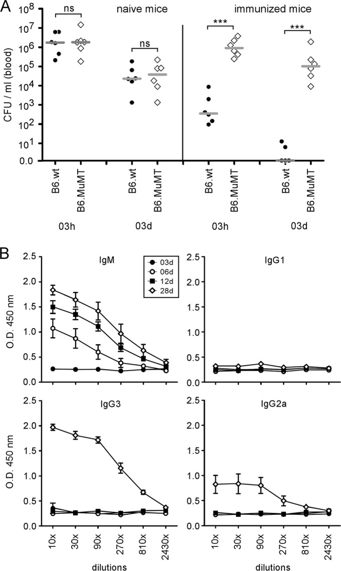
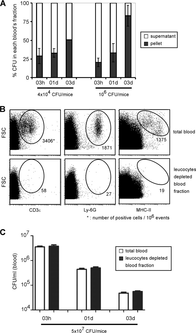
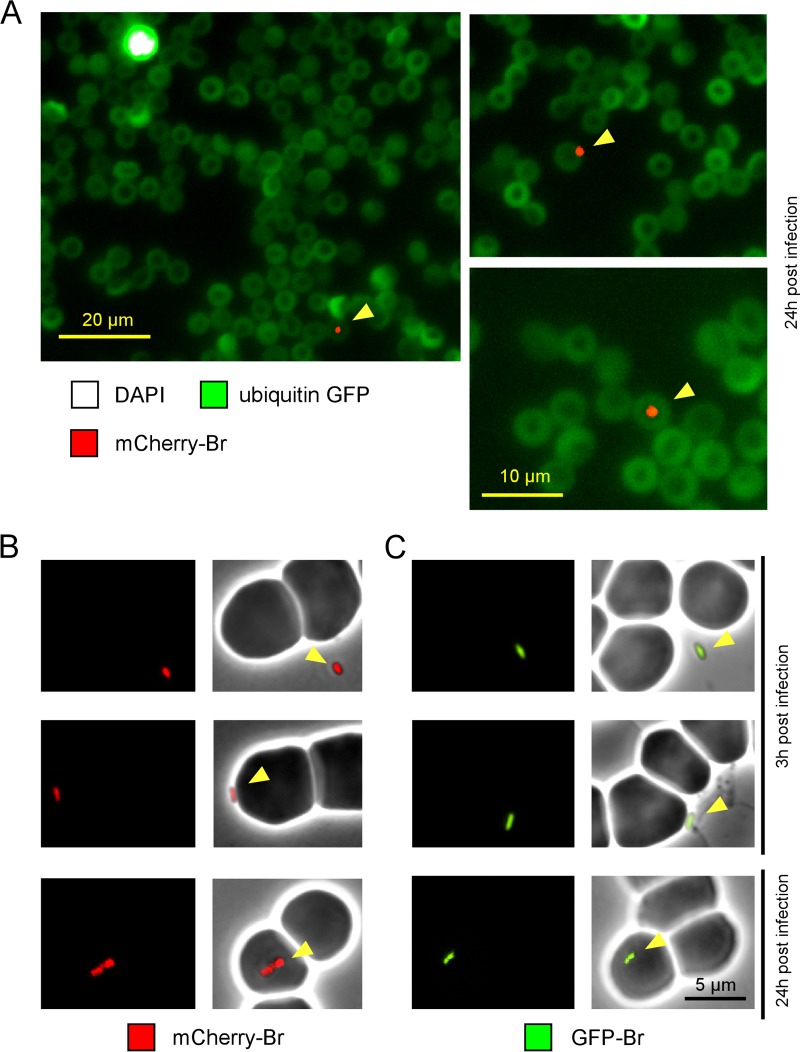
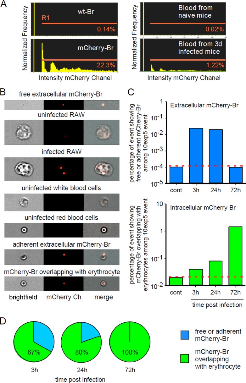
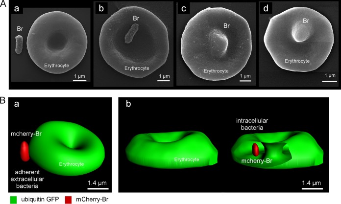
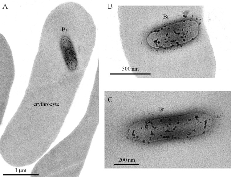
Brucella spp. are facultative intracellular Gram-negative coccobacilli responsible for brucellosis, a worldwide zoonosis. We observed that Brucella melitensis is able to persist for several weeks in the blood of intraperitoneally infected mice and that transferred blood at any time point tested is able to induce infection in naive recipient mice. Bacterial persistence in the blood is dramatically impaired by specific antibodies induced following Brucella vaccination. In contrast to Bartonella, the type IV secretion system and flagellar expression are not critically required for the persistence of Brucella in blood. ImageStream analysis of blood cells showed that following a brief extracellular phase, Brucella is associated mainly with the erythrocytes. Examination by confocal microscopy and transmission electron microscopy formally demonstrated that B. melitensis is able to invade erythrocytes in vivo. The bacteria do not seem to multiply in erythrocytes and are found free in the cytoplasm. Our results open up new areas for investigation and should serve in the development of novel strategies for the treatment or prophylaxis of brucellosis. Invasion of erythrocytes could potentially protect the bacterial cells from the host's immune response and hamper antibiotic treatment and suggests possible Brucella transmission by bloodsucking insects in nature.


Figures

FIG 1
FIG 1
Persistence of infectious B. melitensis in the blood of mice after i.p. infection. (A and B) Wild-type C57BL/6 mice were injected i.p. with 4 × 10⁴, 106, or 5 × 10⁷ CFU of mCherry-expressing B. melitensis (mCherry-Br). and blood was collected at selected time points. The data represent either the means ± SEM of mCherry-Br CFU per ml of blood (A) or the percentage of mice that still had bacteria in the blood (B). The results are representative of two independent experiments. (C) Wild-type C57BL/6 mice were injected i.p. with 106 CFU of mCherry-Br, and blood was collected at 1 (01d), 3, 6, and 13 days p.i. Blood was immediately diluted 20× in PBS and transferred i.p. (500 μl) to recipient wild-type C57BL/6 mice. Two weeks after blood transfer, the mice were sacrificed and the spleens were harvested. The data represent the CFU per spleen and are representative of two independent experiments. The gray bars represent the medians.
FIG 2
FIG 2
Type IV secretion system (VirB)-deficient and flagellum (FliC)-deficient strains of B. melitensis are able to persist in the blood of infected mice until day 3. Wild-type C57BL/6 mice were injected i.p. with 106 CFU of the wild-type (wt), FliC, or VirB strain of B. melitensis, and blood was collected at the indicated time points. The data represent the CFU per ml of blood of individual mice and are representative of three independent experiments. The gray bars represent the medians. **, P < 0.01.
FIG 3
FIG 3
Natural antibodies do not prevent dissemination of B. melitensis in the blood, whereas vaccine-induced specific antibodies reduce the bacteremia following a secondary infection. (A) Wild-type and MuMT−/− C57BL/6 mice were immunized i.p. with 4 × 10⁴ CFU of B. melitensis (vaccinated group) or injected i.p. with PBS (naive group) and then treated with antibiotics for 3 weeks, as described in Materials and Methods. Sixty days after immunization, all mice were injected i.p. with 5 × 10⁷ CFU of mCherry-Br, and blood was collected at the indicated time points. The data represent the CFU per ml of blood and are representative of three independent experiments. The gray bars represent the medians. ***, P < 0.001; ns, not significant. (B) Wild-type C57BL/6 mice were infected with 4 × 10⁴ CFU of B. melitensis. Serum was collected at the indicated time points, and ELISA was performed to determine the isotype distribution of the Brucella-specific antibodies. The data represent the means ± SEM of results for 6 mice. O.D., optical density.
FIG 4
FIG 4
Blood location of B. melitensis after i.p. infection. (A) Wild-type C57BL/6 mice were injected i.p. with 4 × 10⁴ or 106 CFU of mCherry-Br, and blood was collected at the indicated time points. Samples were diluted and centrifuged to separate blood cells from plasma. The data represent the mean ± SD of the percentage of CFU in each blood fraction and are representative of two independent experiments. (B and C) Wild-type C57BL/6 mice were injected i.p. with 5 × 10⁷ CFU of mCherry-Br, and blood was collected at the indicated time points. Samples were diluted and immediately filtered with Plasmodipur filters. (B) Total blood cells and purified erythrocytes were analyzed by flow cytometry using anti-CD3ε, anti-Ly-6G, and anti-MHC-II antibodies in order to confirm the elimination of all leukocytes. (C) The data represent the CFU per ml of total blood or purified erythrocytes and are representative of two independent experiments with 3 mice per group.
FIG 5
FIG 5
Immunofluorescence microscopy of fixed or live bacteria. (A) Ubiquitin-GFP C57BL/6 mice were infected i.p. with 5 × 10⁷ CFU of mCherry-Br and bled at 3 h and 24 h p.i. The blood was fixed and then smeared on slides. The slides were dried overnight and then mounted in Fluoro-Gel medium containing DAPI nucleic acid stain (Electron Microscopy Sciences, Hatfield, PA) and examined under a fluorescence microscope. Brucella was found associated with erythrocytes mainly at 24 h, and thus we present only images starting at 24 h p.i. The figure shows examples of mCherry bacteria associated with DAPI- erythrocytes. The panels are color coded according to the antigen examined. Scale bar = 20 or 10 μm, as indicated. The data are representative of at least 3 independent experiments with a total of more 200 bacteria observed. (B and C) Wild-type C57BL/6 mice were infected with 5 × 10⁷ CFU of mCherry-Br (B) or GFP-Br (C). Blood samples were harvested at 3 h and 24 h p.i. and immediately diluted 10× in PBS. Freshly harvested blood samples were directly dropped on an agarose pad and sealed with VALAP before observation under a fluorescence microscope in a biosafety level III laboratory facility. The data are representative of at least 3 independent experiments, with a total of more than 30 bacteria observed under each condition.
FIG 6
FIG 6
ImageStream analysis of blood from infected mice. Wild-type C57BL/6 mice were injected i.p. with 5 × 10⁷ CFU of mCherry-Br, and blood was collected at 3 h, 24 h, and 72 h p.i. The blood samples were immediately fixed by 3-fold dilution in 4% paraformaldehyde and then analyzed by multispectral imaging flow cytometry (ImageStream 100). As controls, we used a fresh culture of wild-type Brucella, mCherry-Br, and a macrophage cell line (RAW cells) at 3 h post-in vitro infection with mCherry-Br. (A) Comparison of the percentage of events displaying a positive mCherry (channel 4) fluorescent signal (gate R1) in blood from control and infected mice. (B) Representative images of free bacteria and uninfected and infected cells in channels 2 and 4. (C and D) Kinetic analysis of the percentages of free or adherent bacteria (in blue) and bacteria overlapping with cells (in green) found in blood samples from control and infected mice. The location of bacteria was defined by erosion mask. These data are representative of three independent experiments.
FIG 7
FIG 7
Brucella is found associated with or inside erythrocytes by scanning electron and confocal microscopy. (A) Scanning electron microscopy images from blood collected in mice 3 h p.i. by i.p. injection of 5 × 10⁷ CFU of Brucella. Extracellular bacteria (a), attachment of Brucella to erythrocytes (b), and deformed erythrocytes, suggesting Brucella infection (c and d), are shown. (B) Confocal microscopy images of blood collected in mice 24 h p.i. by i.p. injection of 5 × 10⁷ CFU of Brucella. Attachment of Brucella to erythrocytes (a) and Brucella inside erythrocytes (b) are shown.
FIG 8
FIG 8
Brucella is not surrounded by a vacuole in erythrocytes. Transmission electron microscopy analysis of erythrocytes from mice 24 h post-infection by i.p. injection of 5 × 10⁷ CFU of Brucella.

Similar articles

Cited by

References

    1. Godfroid J, Cloeckaert A, Liautard JP, Kohler S, Fretin D, Walravens K, Garin-Bastuji B, Letesson JJ. 2005. From the discovery of the Malta fever's agent to the discovery of a marine mammal reservoir, brucellosis has continuously been a re-emerging zoonosis. Vet. Res. 36:313–326. 10.1051/vetres:2005003 - DOI - PubMed
    1. Cutler SJ, Whatmore AM, Commander NJ. 2005. Brucellosis—new aspects of an old disease. J. Appl. Microbiol. 98:1270–1281. 10.1111/j.1365-2672.2005.02622.x - DOI - PubMed
    1. Pappas G, Papadimitriou P, Akritidis N, Christou L, Tsianos EV. 2006. The new global map of human brucellosis. Lancet Infect. Dis. 6:91–99. 10.1016/S1473-3099(06)70382-6 - DOI - PubMed
    1. Gregoire F, Mousset B, Hanrez D, Michaux C, Walravens K, Linden A. 2012. A serological and bacteriological survey of brucellosis in wild boar (Sus scrofa) in Belgium. BMC Vet. Res. 8:80. 10.1186/1746-6148-8-80 - DOI - PMC - PubMed
    1. Zheludkov MM, Tsirel'son LE. 2010. Reservoirs of Brucella infection in nature. Biol. Bull. 37:709–715. 10.1134/S106235901007006X - DOI

Publication types

Substances

LinkOut - more resources