Review
. 2014 Sep;15(9):437.
doi: 10.1007/s11934-014-0437-y.

Confocal laser endomicroscopy of bladder and upper tract urothelial carcinoma: a new era of optical diagnosis?

Affiliations
Review

Confocal laser endomicroscopy of bladder and upper tract urothelial carcinoma: a new era of optical diagnosis?

Stephanie P Chen et al. Curr Urol Rep. 2014 Sep.

Abstract

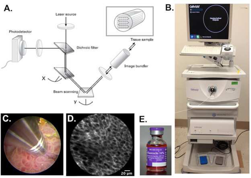
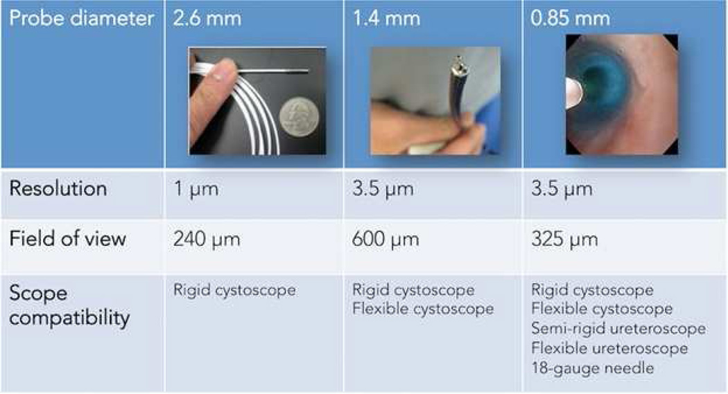
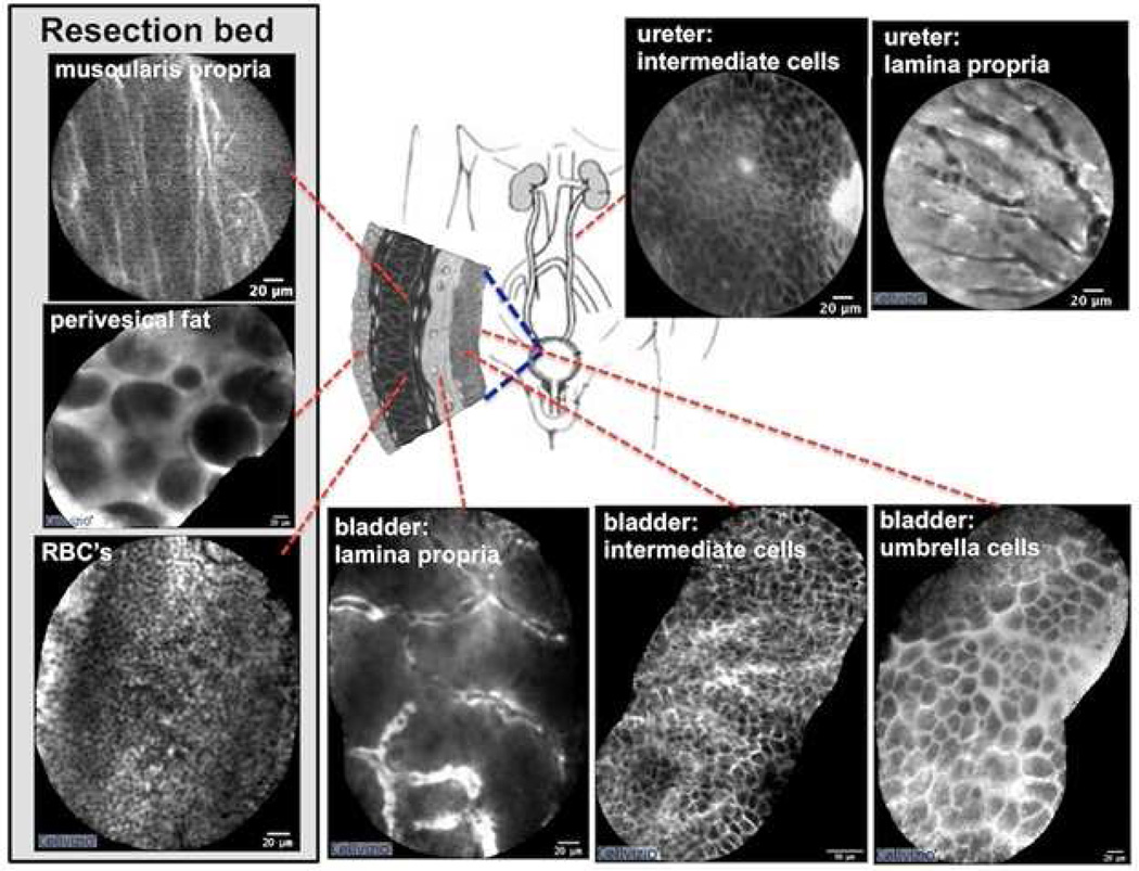
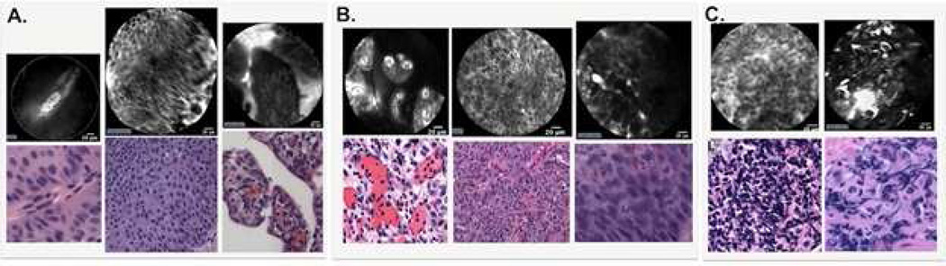
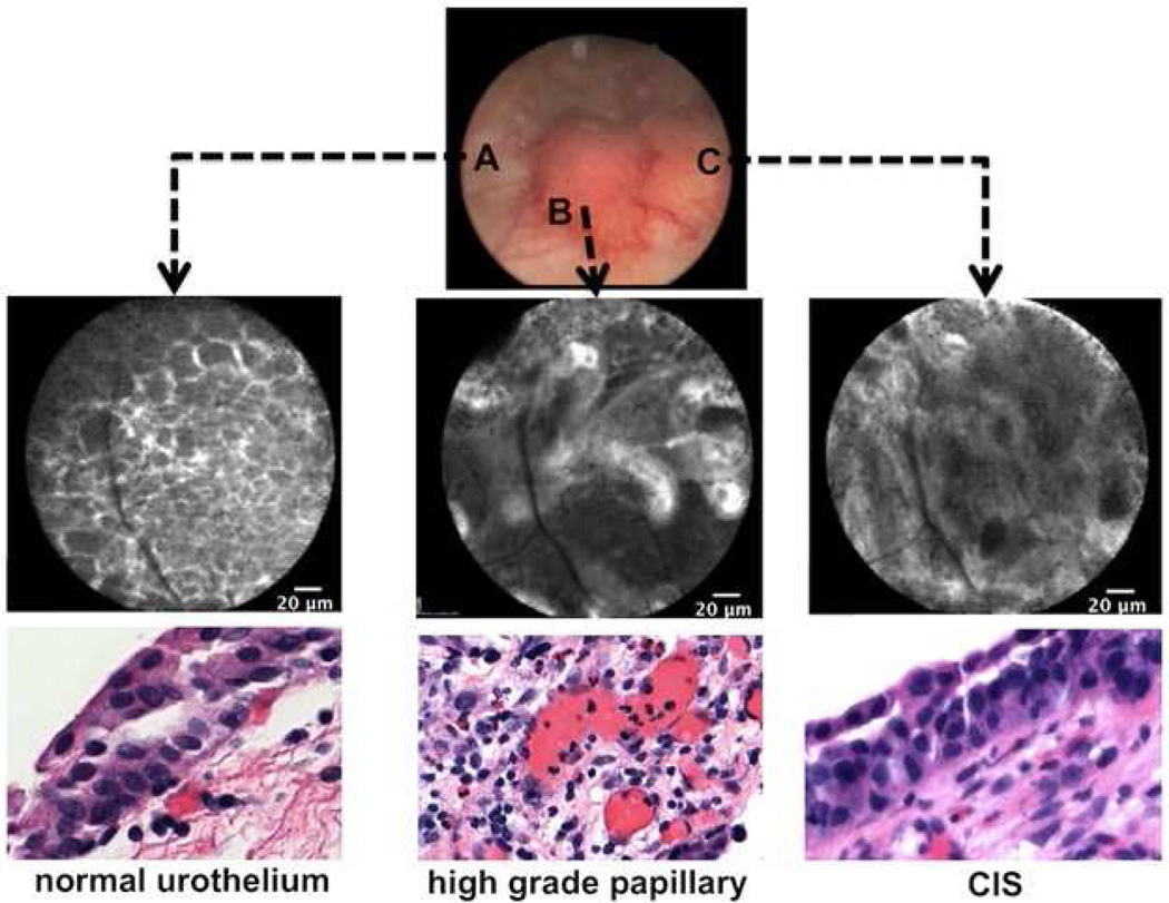
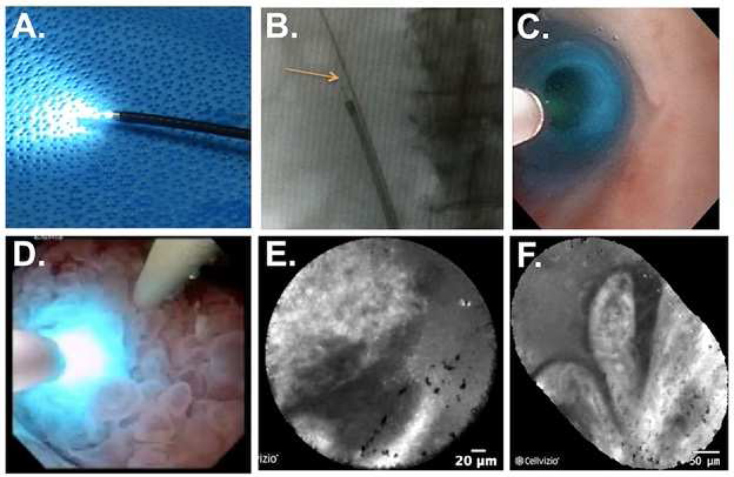
Urothelial carcinoma of the bladder and upper tract pose significant diagnostic and therapeutic challenges. White light endoscopy plays a central role in the management of urothelial carcinoma but has several well-recognized shortcomings. New optical imaging technologies may improve diagnostic accuracy, enhance local cancer control, and better stratify treatment options. Confocal laser endomicroscopy enables dynamic imaging of the cellular structures below the mucosal surface and holds promise in providing real time optical diagnosis and grading of urothelial carcinoma. A variety of imaging probes are available that are compatible with the full spectrum of cystoscopes and ureteroscopes. We review the underlying principles and technique of confocal laser endomicroscopy in the urinary tract, with emphasis on specific application towards urothelial carcinoma. While the available data are largely related to urothelial carcinoma of the bladder, the lessons learned are directly applicable to the upper tract, where the clinical needs are significant. Ongoing efforts to optimize this technology offer an exciting glimpse into future advances in optical imaging and intraoperative image guidance.


Conflict of interest statement

Conflict of Interest

Dr. Stephanie P. Chen and Dr. Joseph C. Liao each declare no potential conflicts of interest.

Figures

Fig. 1
Fig. 1
Mechanism and method of confocal laser endomicroscopy. A) Schematic representation of the technology underlying CLE. A laser beam is directed to the tissue sample via an image bundler. The resulting emission of light is retrieved and filtered through a pinhole before reaching the photodetector; B) Clinical grade CLE system (Cellvizio) from Mauna Kea Technologies. The workstation includes a laser scanning unit for probe attachment and a computer with software for image acquisition and processing. C) Direct probe contact of a papillary tumor for image acquisition. D) High-resolution image of subsurface cellular features acquired from CLE. The fluorescence signal is coming from the extracellular matrix stained by fluorescein. E) Fluorescein, an FDA-approved contrast agent, can be administered intravenously or topically for CLE imaging
Fig. 2
Fig. 2
Comparison of available probes for confocal laser endomicroscopy. Probes range in diameter from 2.6 mm to 0.85 mm. Decreasing spatial resolution by downsizing the probes is offset by greater compatibility with a larger array of endoscopes, including flexible scopes. The field of view is most narrow with the 2.6 mm probe and widest with the 1.4 mm probe, with the 0.85 mm probe falling in between
Fig. 3
Fig. 3
Confocal images of normal anatomic landmarks within the urinary tract. Normal urothelium is characterized by superficial polygonal-shaped umbrella cells and underlying intermediate cells. The lamina propria is characterized by a vacular network with a relatively acellular matrix. Characteristic findings of the tumor resection bed include multi-directional fibers consistent with the muscularis propria, large adipocytes within perivesical fat, and copious red blood cells. The resection bed was imaged after the tumor resection. In the upper urinary tract, images of the urothelium are similar to the lower tract. Images of the lower urinary tract were acquired in vivo while those of the upper urinary tract were obtained ex vivo. Video mosaicing was utilized to expand the field of view for select images
Fig. 4
Fig. 4
Diagnostic imaging criteria for urothelial carcinoma. A) Low-grade cancer are characterized by organized, densely-packed, monomorphic cells, the absence of umbrella cells, papillary structures, and fibrovascular stalks. B) High-grade cancer show disorganized, pleomorphic cells, indistinct borders with loss of cellular cohesion, the absence of umbrella cells, and fibrovascular stalks with distorted vasculature. C) Benign, inflammatory lesions feature small, infiltrative, monomorphic cells in the lamina propria loosely arranged and absent fibrovascular stalks. Normal urothelium is described in Fig. 3. Using confocal laser endomicroscopy adjunct to white light cystoscopy, these diagnostic imaging criteria demonstrated substantial agreement among urologists familiar with the technology (ĸ 0.80) and moderate agreement among novice users (ĸ 0.59). Sensitivity and specificity of the diagnostic criteria were reported as 89 % and 88 %, respectively
Fig. 5
Fig. 5
Intraoperative image guidance of a tumor seen under white light with corresponding confocal imaging and histology. A) Normal urothelium surrounding the papillary tumor highlighting organized, monomorphic cells. B) High-grade, papillary tumor with papillary structures and distorted microvasculature. C) High-grade, carcinoma-in-situ featuring pleomorphic cells and disorganized microarchitecture
Fig. 6
Fig. 6
Application of confocal laser endomicroscopy to the upper urinary tract. A) A 0.85 mm probe inserted into a flexible ureteroscope. B) Fluoroscopy image of the flexible ureteroscope in the ureter. The arrow is pointing to the confocal probe fitted through the ureteroscope. C) Ureteroscopic view of the confocal probe inside a normal ureter. D) Confocal probe in direct contact with a large, papillary tumor in the renal pelvis. E) CLE image of tumor in D showing the papillary border. F) Mosaic image of the papillary tumor showing the papillary structure of the tumor shown in D. Fine streaks seen in the papillary structures represent the fibrovascular stalk present in cancerous lesions

Similar articles

Cited by

References

    1. U.S. Cancer Statistics Working Group. U.S. Department of Health and Human Services, Centers for Disease Control and Prevention and National Cancer Institute; 2013. United States Cancer Statistics, 1999–2010 Incidence and Mortality Web-based Report.
    1. Siegel R, Ma J, Zou Z, Jemal A. Cancer statistics, 2014. CA. Cancer J. Clin. 2014;64:9–29. - PubMed
    1. Kirkali Z, et al. Bladder cancer: epidemiology, staging and grading, and diagnosis. Urology. 2005;66:4–34. - PubMed
    1. Morgan TM, Clark PE. Bladder cancer. Curr. Opin. Oncol. 2010;22:242–249. - PubMed
    1. Sylvester RJ, et al. Predicting recurrence and progression in individual patients with stage Ta T1 bladder cancer using EORTC risk tables: a combined analysis of 2596 patients from seven EORTC trials. Eur. Urol. 2006;49:466–465. discussion 475–477. - PubMed

Publication types

MeSH terms

LinkOut - more resources