Review
. 2014:68:493-520.
doi: 10.1146/annurev-micro-091313-103550. Epub 2014 Jun 16.

Regulation of virulence of Entamoeba histolytica

Affiliations
Review

Regulation of virulence of Entamoeba histolytica

Chelsea Marie et al. Annu Rev Microbiol. 2014.

Abstract

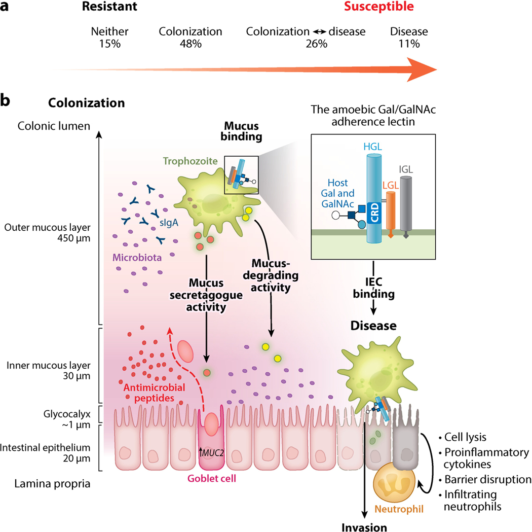
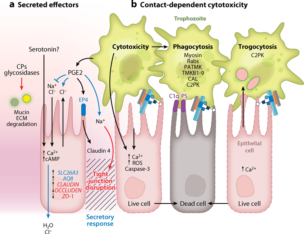
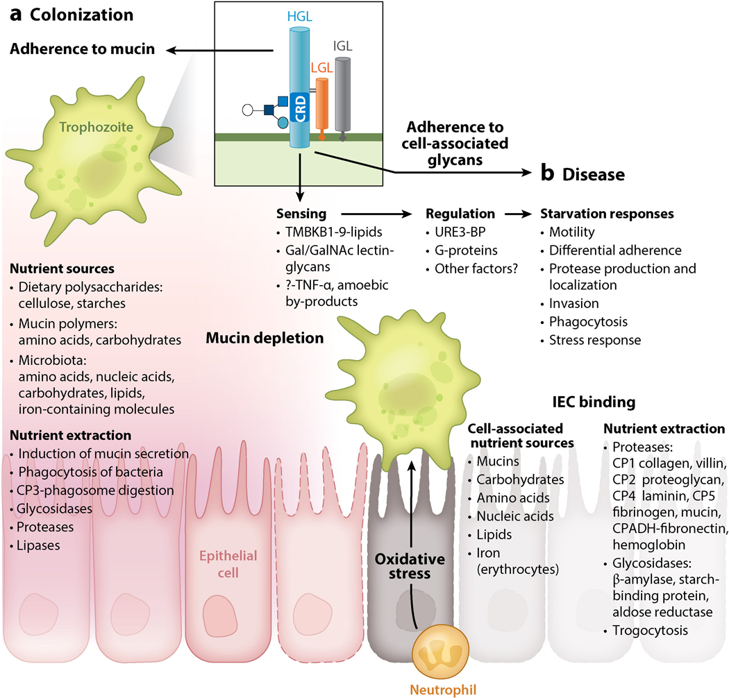
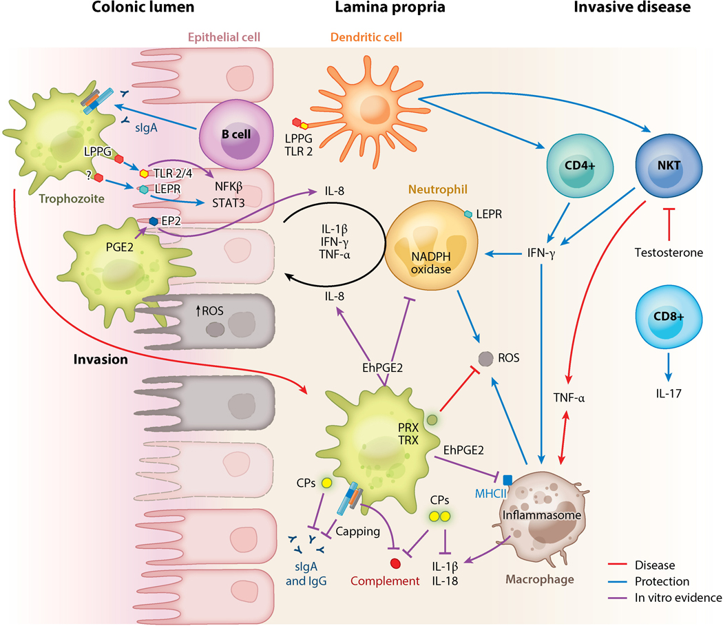
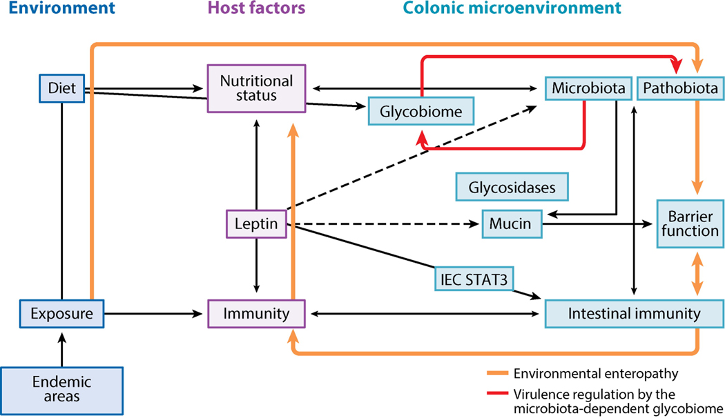
Entamoeba histolytica is the third-leading cause of parasitic mortality globally. E. histolytica infection generally does not cause symptoms, but the parasite has potent pathogenic potential. The origins, benefits, and triggers of amoebic virulence are complex. Amoebic pathogenesis entails depletion of the host mucosal barrier, adherence to the colonic lumen, cytotoxicity, and invasion of the colonic epithelium. Parasite damage results in colitis and, in some cases, disseminated disease. Both host and parasite genotypes influence the development of disease, as do the regulatory responses they govern at the host-pathogen interface. Host environmental factors determine parasite transmission and shape the colonic microenvironment E. histolytica infects. Here we highlight research that illuminates novel links between host, parasite, and environmental factors in the regulation of E. histolytica virulence.

Keywords: carbohydrate utilization; microbiota; mucus; pathobiota; virulence.


Figures

Figure 1
Figure 1
E. histolytica virulence depends on a dynamic interaction in the infected host. (a) Continuum of E. histolytica disease in a natural population. Analysis of diarrheal and monthly surveillance stool samples for E. histolytica detected four possible outcomes in the first two years of life: (1) no evidence of infection, (2) colonization with no E. histolytica–associated diarrhea, (3) diarrhea with prior asymptomatic colonization and/or subsequent asymptomatic persistence, or (4) E. histolytica–associated diarrhea with no previous colonization. This pattern reinforces the importance of both parasite and host factors in the outcome of an E. histolytica infection. (b) To establish infection, E. histolytica must bind and adhere in the host colon. Adherence is mediated by an amoebic lectin with a carbohydrate-recognition domain (CRD) that binds galactose (Gal) and N-acetyl-d-galactosamine (GalNAc) on host glycoconjugates with high affinity. The Gal/GalNAc lectin is composed of heavy (HGL), intermediate (IGL), and light (LGL) subunits. The CRD is located on the HGL, which also contains a putative intracellular signaling domain. HGL forms a disulfide bond with LGL. The HGL-LGL heterodimer can associate with the glycosylphosphatidylinositol-anchored IGL, but this subunit does not have a well-defined function (inset). Colonic mucin forms a dense polymeric gel over the intestinal epithelium, which trophozoites bind to with high affinity. Trophozoites also induce mucin secretion by goblet cells. In colonization, mucin binding mediates attachment and provides a nutrient source for E. histolytica. Mucin polymers may be degraded by amoebic proteases and glycosidases for nutrients, and the mucosal microbiota provides a nutrient source via amoebic phagocytosis. The transition from colonization to disease is marked by destruction of the mucin barrier. Mucus depletion may result from enhanced amoebic degradation and/or depletion of mucin stores by continual secretion during chronic infection. Other factors including coinfections, host diet, and disruption of the microbiota can also mediate mucus depletion. Mucus depletion exposes the intestinal epithelium to E. histolytica trophozoites. The amoebic lectin CRD binds to Gal and GalNAc on exposed intestinal epithelial cells (IECs) and the cell-associated glycocalyx. Adherence to IECs results in amoebic cytotoxicity and the release of proinflammatory molecules. Abbreviation: sIgA, secretory immunoglobulin A.
Figure 2
Figure 2
Pathology at the intestinal epithelium. (a) Physiological mediators of E. histolytica diarrhea. Secreted amoebic effectors have contact-independent physiological effects on intestinal epithelial cells. (1) Proteases and glycosidases degrade mucus and extracellular matrix (ECM) proteins. (2) Amoebic PG2 disrupts barrier function by binding to host EP4, leading to altered expression and localization of tight junction proteins including zona occludens proteins and claudins. (3) PG2 also increased secretion and disrupted ion gradients, leading to decreased cellular Na2+ absorption and increased Na2+ and Cl− secretion at the apical surface. Amoebic serotonin is present in amoebic lysates, but it is not known if it is secreted. Serotonin elevates intracellular Ca2+ and cAMP, leading to increased H2O and Cl− secretion at the serosal surface. Disruption of barrier function further disrupts ion gradients at the intestinal epithelium, and these effects are likely the physiological mediators of amoebic diarrhea (secretory response, blue; tight junction disruption, red). (b) In vitro trophozoites must adhere to target cells to induce death. Contact-dependent killing can be mediated by amoebic activation of host caspase-3 through an undefined mechanism and a rapid apoptotic-like death, preceded by elevated intracellular Ca2+ and reactive oxygen species. E. histolytica phagocytosis is initiated by exposed C1q and phosphatidylserine (PS) on apoptotic cells, which are bound by amoebic calreticulin (CAL) and C2K, respectively. Amoebic kinases PATMK, TMKB1–9, and TMK39 are also involved in phagocytosis. Phagosome formation requires vesicular trafficking and cytoskeletal rearrangement controlled by G-proteins and amoebic myosin. Trogocytosis is a distinct contact-dependent mechanism of amoebic cytotoxicity. In trogocytosis, trophozoites actively ingest pieces of living cells, resulting in membrane disruption and rapid target cell death. Trogocytosis also requires amoebic C2PK and leads to increased intracellular Ca2+ prior to cell death; however, in trogocytosis E. histolytica does not ingest cells after killing. Other abbreviations: CP, cysteine protease; PGE2, prostaglandin 2; ROS, reactive oxygen species.
Figure 3
Figure 3
E. histolytica adaptive ability mediates nutrient extraction and survival in the fluctuating colonic environment. (a) Colonic nutrient sources include dietary polysaccharides, microbiota, and human cellular molecules including mucin. Microbial glycosidases degrade complex polysaccharides into fatty acids absorbed by intestinal epithelial cells (IECs) and sugars for their own metabolism. An intact mucous layer provides plentiful nutrients from mucin and microbiota, inducing a parasite program for colonization. (b) Upon mucus depletion, nutrient starvation induces virulence and activates the transcriptional regulator URE3-BP. Starvation responses include decreased adherence and enhanced motility and oxidative stress resistance. Upon adherence to host cells, virulence factors are induced to enable extraction of cell-associated nutrients. Abbreviations: CP, cysteine protease; CRD, carbohydrate-recognition domain; Gal, galactose; GalNAc, N-acetyl-d-galactosamine; HGL, heavy lectin subunit; IGL, intermediate lectin subunit; LGL, light lectin subunit.
Figure 4
Figure 4
Immune regulators of E. histolytica virulence. The mucin barrier, mucosal sIgA to the carbohydrate-recognition domain of the galactose (Gal)/N-acetyl-d-galactosamine (GalNAc) lectin, and leptin signaling via a leptin receptor (LEPR) at the epithelium are critical determinates of protection in the colonic lumen. The Gal/GalNAc lectin activates Toll-like receptors (TLRs) on intestinal epithelial cells (IECs), leading to IL-8 secretion and neutrophil recruitment. IEC damage from amoebic cytotoxicity further induces secretion of proinflammatory mediators and disrupts tight junctions, enhancing neutrophil infiltration. Neutrophil reactive oxygen species (ROS) can kill trophozoites; however, trophozoites also kill immune cells. ROS can also exacerbate host tissue damage. Trophozoite peroxiredoxin (PRX) and thioredoxin (TRX) detoxify ROS. Amoebic PG2 can suppress ROS production and impair major histocompatibility complex II (MHCII) expression on macrophages, inhibiting their antigen-presenting ability. Dendritic cells (DCs) in the lamina propria also act as antigen-presenting cells and recognize amoebic LPPG via TLR-2. DCs can activate natural killer and CD+ T cells. In invasive disease IFN-γ-producing natural killer T cells (NKTs) are associated with production. TNF-α production by NKTs and macrophages is associated with increased disease severity. The Gal/GalNAc lectin activates the NLRP3 inflammasome and secretion of IL-1β and IL-18 in macrophages in vitro, though it is not known if this is protective or deleterious. Amoebic proteases cleave complement, IgA, IgG, pro-IL-1β, and IL-18. The Gal/GalNAc lectin inhibits formation of complement membrane attack complex (MAC) and can mediate the capping and shedding of bound antibodies. Processes associated with protection in vivo (animals and/or humans) are indicated with blue arrows; processes associated with disease in vivo are shown in red. Purple lines indicate in vitro evidence. Other abbreviations: CP, cysteine protease; PGE2, prostaglandin 2; sIgA, secretory immunoglobulin A.
Figure 5
Figure 5
The vicious cycle of enteric infection and malnutrition. E. histolytica cysts enter the host via fecally contaminated food and water. In areas where E. histolytica infection is endemic, amoebic infection is pervasive and accompanied by multiple other enteric pathogens. The linked immune and nutritional status of the host determines whether infections will be resolved or established in the host intestine. Leptin levels regulate the immunodeficiency of malnutrition, and reduced leptin increases susceptibility to E. histolytica and other pathogens. Leptin signaling is critical for protection from E. histolytica at the intestinal epithelium. In the absence of immune clearance or treatment, E. histolytica and other enteric pathogens establish chronic infection as part of the pathobiota. The pathobiota causes chronic intestinal inflammation and mucus depletion. Chronic inflammatory responses disrupt the absorptive and barrier functions of the intestine, worsening malnutrition and leading to environmental enteropathy. E. histolytica causes dysbiosis with potential consequences for host nutrition and immunity, as the microbiota mediates intestinal immune homeostasis and nutrient extraction. The microbiota stimulates antimicrobial peptide and mucin production by intestinal epithelial cells (IECs), leading to exclusion of pathogens. Microbial metabolism of dietary and host-derived carbohydrates is essential for host nutrient absorption and for microbial metabolism and leads to competitive exclusion of some enteric pathogens. Microbial metabolism of complex polysaccharides in the colon produces short-chain fatty acids and oligosaccharides, which are critical for host nutrition. E. histolytica encodes sugar transporters and glycosidase genes from prokaryotes, indicating that E. histolytica is capable of exploiting free sugars produced by microbial metabolism. The microbially derived glycobiome is also an emerging regulator of enteric pathogen virulence. Dashed lines are hypothetical.

Similar articles

Cited by

References

    1. Abd-Alla MD, Jackson TFGH, Rogers T, Reddy S, Ravdin JI. 2006. Mucosal immunity to asymptomatic entamoeba histolytica and entamoeba dispar infection is associated with a peak intestinal anti-lectin immunoglobulin a antibody response. Infect. Immun. 74(7):3897–3903 - PMC - PubMed
    1. Abhyankar MM, Shrimal S, Gilchrist CA, Bhattacharya A, Petri WA Jr. 2012. The entamoeba histolytica serum-inducible transmembrane kinase ehtmkb1–9 is involved in intestinal amebiasis. Int. J. Parasitol. Drugs Drug Resist. 2:243–48 - PMC - PubMed
    1. Ackers J, Clark G, Diamond L, Duchene M, Cantellano M, et al. 1997. Who/paho/unesco report of a consultation of experts on amoebiasis
    1. Ali IKM, Clark CG, Petri WA Jr. 2008. Molecular epidemiology of amebiasis. Infect. Genet. Evol. J. Mol. Epidemiol. Evol. Genet. Infect. Dis. 8(5):698–707 - PMC - PubMed
    1. Ali IKM, Haque R, Alam F, Kabir M, Siddique A, Petri WA Jr. 2012. Evidence for a link between locus r-r sequence type and outcome of infection with entamoeba histolytica. Clin. Microbiol. Infect. Off. Publ. Eur. Soc. Clin. Microbiol. Infect. Dis. 18(7):E235–237 - PMC - PubMed

Publication types

Substances

LinkOut - more resources