. 2014 Oct;28(10):4347-58.
doi: 10.1096/fj.14-251611. Epub 2014 Jul 1.

Role of sphingosine kinase 1 and sphingosine-1-phosphate in CD40 signaling and IgE class switching

Affiliations

Role of sphingosine kinase 1 and sphingosine-1-phosphate in CD40 signaling and IgE class switching

Eugene Y Kim et al. FASEB J. 2014 Oct.

Abstract

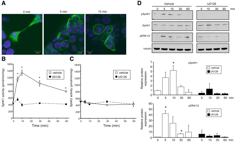
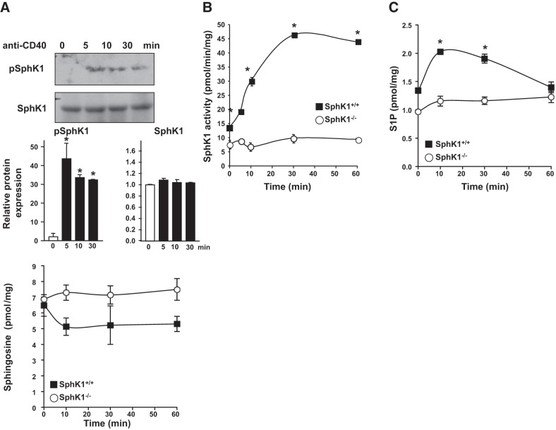
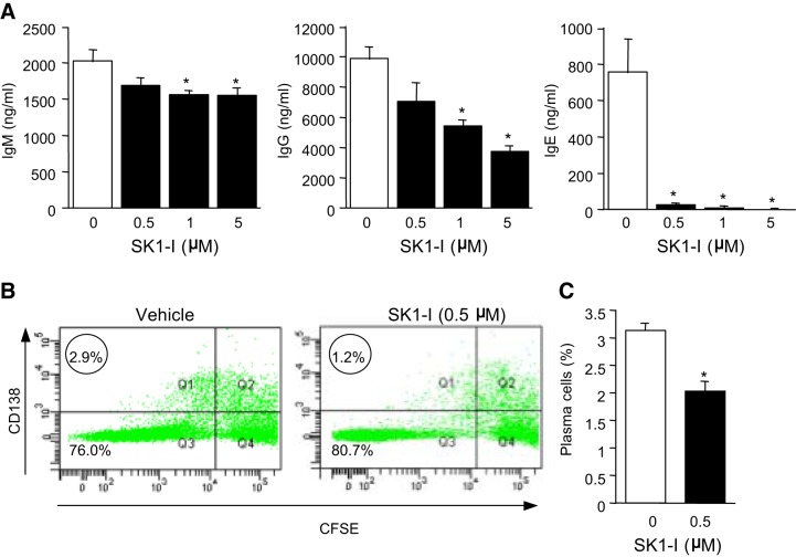
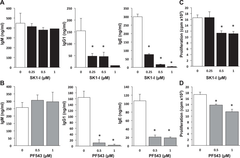
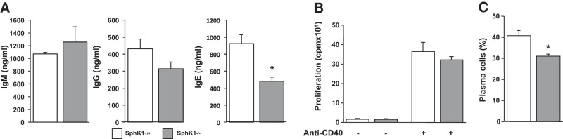
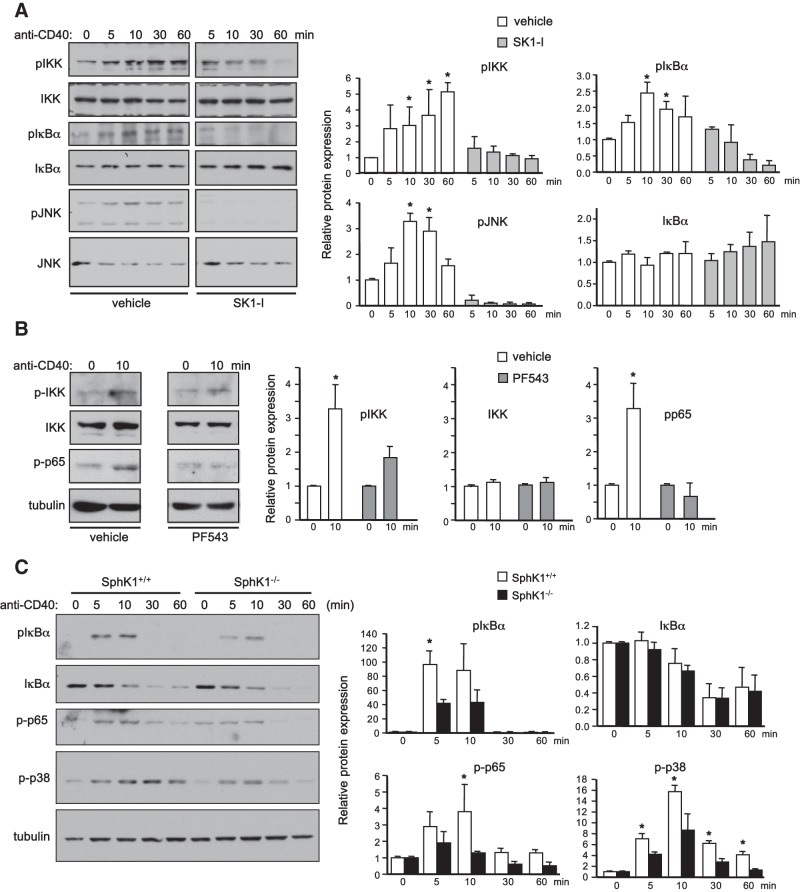
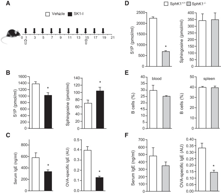
The tumor necrosis factor (TNF) receptor family member CD40 plays an essential role in the activation of antigen-presenting cells, B cell maturation, and immunoglobulin (Ig) class switching critical for adaptive immunity. Although the bioactive sphingolipid metabolite sphingosine-1-phosphate (S1P) and the kinase that produces it, sphingosine kinase 1 (SphK1), have long been implicated in the actions of TNF mediated by engagement of TNFR1, nothing is yet known of their role in CD40-mediated events. We have now found that ligation of CD40 activates and translocates SphK1 to the plasma membrane, leading to generation of S1P. SphK1 inhibition in human tonsil B cells, as well as inhibition or deletion of SphK1 in mouse splenic B cells, significantly reduced CD40-mediated Ig class switching and plasma cell differentiation ex vivo. Optimal activation of downstream CD40 signaling pathways, including NF-κB, p38, and JNK, also required SphK1. In mice treated with a SphK1 inhibitor or in SphK1(-/-) mice, isotype switching to antigen-specific IgE was decreased in vivo by 70 and 55%, respectively. Our results indicate that SphK1 is important for CD40-mediated B cell activation and regulation of humoral responses and suggest that targeting SphK1 might be a useful therapeutic approach to control antigen-specific IgE production.

Keywords: B cells; NF-κB; inflammation; sphingolipids.


Figures

Figure 1.
Figure 1.
CD40 ligation translocates and activates SphK1. A) HEK-CD40 cells were transfected with V5-SphK1 and stimulated with agonistic anti-CD40 antibody for the indicated times. Cells were stained with Hoechst (blue) and V5 antibody (green) and visualized by confocal microscopy. Scale bars = 10 μm. B, C) Serum-starved HEK-CD40 cells were stimulated with anti-CD40 antibody for the indicated times in the presence of vehicle or U0126 (1 μM). SphK1 activity (B) and SphK2 activity (C) in cell lysates were measured by formation of S1P. Data are expressed as means ± sd from triplicate determinations. Similar results were obtained in 2 additional experiments. D) In duplicate cultures, phospho-Ser225-SphK1, total SphK1, and pERK1/2 were evaluated by Western blot analysis with the indicated antibodies. One set of representative data is shown from 3 independent experiments. Quantified data by densitometry are expressed as mean ± sd fold vs. time 0; n = 3.
Figure 2.
Figure 2.
CD40 stimulation activates SphK1 and increases S1P levels in B cells. A) Human tonsil B cells were stimulated with anti-CD40 antibody, and levels of phospho-Ser225-SphK1 and total SphK1 were evaluated by Western blot analysis. One set of representative data is shown from 2 independent experiments. Quantified data by densitometry are expressed as mean ± sd fold vs. time 0; n = 2. *P < 0.05 vs. time 0. B) Splenic B cells purified from SphK1+/+ and SphK1−/− mice were stimulated with agonistic CD40 antibody for the indicated times. B) SphK1 activity was measured by formation of [32P]S1P, and data are expressed as means ± sd from triplicate determinations. Where not shown, the size of the symbols indicates the sd. C, D) Mass levels of S1P (C) and sphingosine (D) were measured by LC-ESI-MS/MS. Data are expressed as means ± sd from triplicate determinations. Similar results were obtained in 2 additional independent experiments. *P < 0.05 vs. time 0.
Figure 3.
Figure 3.
Effect of SphK1 inhibition in human B cells on CD40-mediated isotype switching and plasma cell differentiation. Human B cells (A, B) were isolated from tonsils by positive selection using IgD-specific beads by MACS sorting. B cells were cultured with anti-CD40 antibody, IL-4, and IL-21 for 14 d (A) or 7 d (B, C) in the absence or presence of the indicated concentration of SK1-I. A) Antibody levels were measured by antibody-specific ELISAs. Data are expressed as means ± sd from triplicate determinations. Similar results were obtained in 2 additional independent experiments. *P < 0.05. B, C) Plasma cell differentiation was analyzed by FACS. B) Representative flow cytometry plots. Circled numbers indicate frequency of plasma cells (CD138+ and CFSElow). C) Percentage plasma cells from another donor. Data are expressed as means ± sd from triplicate determinations.
Figure 4.
Figure 4.
SphK1 inhibitors suppress mouse B-cell isotype switching ex vivo. Splenic B cells from C57BL/6 mice were isolated by negative selection using anti-CD43 beads by MACS sorting. B cells were cultured with agonistic anti-CD40 antibody and IL-4 for 8 d (A, B) or 3 d (C, D) in the presence of the indicated concentrations of SK1-I (A, C) or PF543 (B, D). A, B) Antibody levels were measured by ELISA. C, D) DNA synthesis was measured by [3H]thymidine uptake. Data are expressed as means ± sd from triplicate determinations. Similar results were obtained in 2 additional experiments. *P < 0.05.
Figure 5.
Figure 5.
SphK1 deletion in B cells impairs differentiation to plasma cells and isotype switching. Splenic B cells from SphK1−/− mice and wild-type littermates were isolated by negative selection using anti-CD43 beads by MACS sorting and cultured with agonistic anti-CD40 antibody and IL-4. A) After 8 d, antibody levels were measured by ELISA. B) After 3 d, DNA synthesis was measured by [3H]thymidine uptake. C) After 7 d, differentiation of B cells to plasma cells was determined by FACS. Data are expressed as means ± sd from triplicate determinations. Similar results were obtained in 2 additional experiments. *P < 0.05.
Figure 6.
Figure 6.
SphK1 is important for optimal CD40 signaling. A, B) Serum-starved HEK-CD40 cells were pretreated with vehicle, SK1-I (2.5 μM), or PF543 (5 μM) for 30 min and then stimulated with anti-CD40 antibody for various times as indicated. C) Splenic B cells purified from SphK1+/+ and SphK1−/− mice were stimulated with anti-CD40 antibody for various times. A–C) Equal amounts of cell lysate proteins were analyzed by Western blotting with the indicated antibodies. One set of representative data is shown from 3 independent experiments. Quantified data by densitometry are expressed as mean ± sd fold vs. time 0; n = 3. *P < 0.05 vs. time 0.
Figure 7.
Figure 7.
SphK1 is involved in antigen-specific isotype switching in vivo. A, B) C57BL/6 mice were immunized with OVA/alum (open arrows) and then treated with saline (vehicle) or SK1-I (10 mg/kg, i.p.) every other day (solid arrows), given an OVA boost on d 15, and euthanized on d 21. B) Blood levels of S1P and sphingosine were determined by LC-ESI-MS/MS. Data are means ± sd; n = 3 mice/group. C) Serum IgE and OVA-specific IgE levels (1:10 dilutions) were determined by ELISA. Data are means ± se; n = 5 mice/group. B, C) Similar results were obtained in 3 independent experiments. *P < 0.05 vs. vehicle treatment. D–F) SphK1+/+ and SphK1−/− mice were immunized with OVA/alum, given an OVA boost on d 15, and euthanized on d 21. D) Blood levels of S1P and sphingosine of SphK1+/+ and SphK1−/− mice were determined by LC-ESI-MS/MS. E) FACS analysis of B220+ B cells in blood and spleen of SphK1+/+ and SphK1−/− mice. F) Serum IgE and OVA-specific IgE levels (1:10 dilutions) were determined by ELISA. Data are representative of 3 independent experiments and are means ± se; n = 5 mice/group. *P < 0.05 vs. WT mice.

References

    1. Van Kooten C., Banchereau J. (2000) CD40-CD40 ligand. J. Leukoc. Biol. 67, 2–17 - PubMed
    1. Bishop G. A. (2004) The multifaceted roles of TRAFs in the regulation of B-cell function. Nat. Rev. Immunol. 4, 775–786 - PubMed
    1. Rickert R. C., Jellusova J., Miletic A. V. (2011) Signaling by the tumor necrosis factor receptor superfamily in B-cell biology and disease. Immunol. Rev. 244, 115–133 - PMC - PubMed
    1. Kawabe T., Naka T., Yoshida K., Tanaka T., Fujiwara H., Suematsu S., Yoshida N., Kishimoto T., Kikutani H. (1994) The immune responses in CD40-deficient mice: impaired immunoglobulin class switching and germinal center formation. Immunity 1, 167–178 - PubMed
    1. Jabara H. H., Fu S. M., Geha R. S., Vercelli D. (1990) CD40 and IgE: synergism between anti-CD40 monoclonal antibody and interleukin 4 in the induction of IgE synthesis by highly purified human B cells. J. Exp. Med. 172, 1861–1864 - PMC - PubMed

Publication types

MeSH terms