. 2014 Jul 7:14:226.
doi: 10.1186/1472-6882-14-226.

In vitro cytotoxicity of Artemisia vulgaris L. essential oil is mediated by a mitochondria-dependent apoptosis in HL-60 leukemic cell line

Affiliations

In vitro cytotoxicity of Artemisia vulgaris L. essential oil is mediated by a mitochondria-dependent apoptosis in HL-60 leukemic cell line

Ayman M Saleh et al. BMC Complement Altern Med. .

Abstract

Background: The essential oil (EO) of Artemisia vulgaris L. has been traditionally used worldwide for treating a large number of diseases. Although major components in A. vulgaris EO have been shown to inhibit growth of different cancer cells, as pure compounds or part of other plants extracted oil, no information is known about its anti-proliferative activities. Therefore, the current investigation has evaluated the toxicity of the plant extracted oil from buds (AVO-b) and leaves (AVO-l) and characterized their growth inhibitory effects on cancer cells.

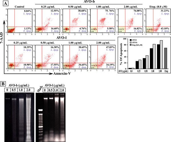
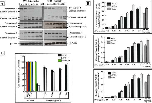
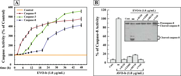
Methods: AVO-b and AVO-l from A. vulgaris L. were extracted by hydrodistillation, and their effect on the viability of human HL-60 promyelocytic leukemia and various other cancer cell lines was tested using MTT assay. Flow cytometric analysis of apoptosis, DNA fragmentation assay, caspases enzymatic activities and Western blotting were used to determine the apoptotic pathway triggered by their action on HL-60 cells.

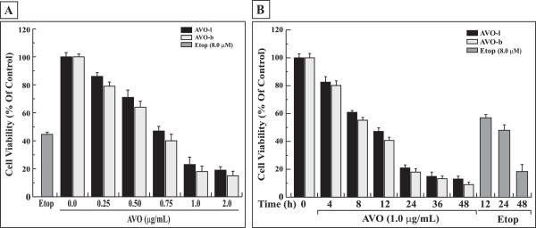
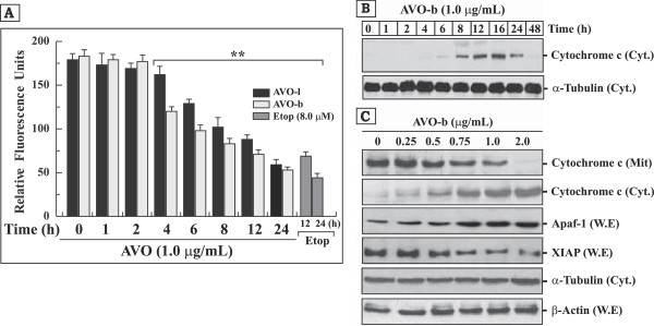
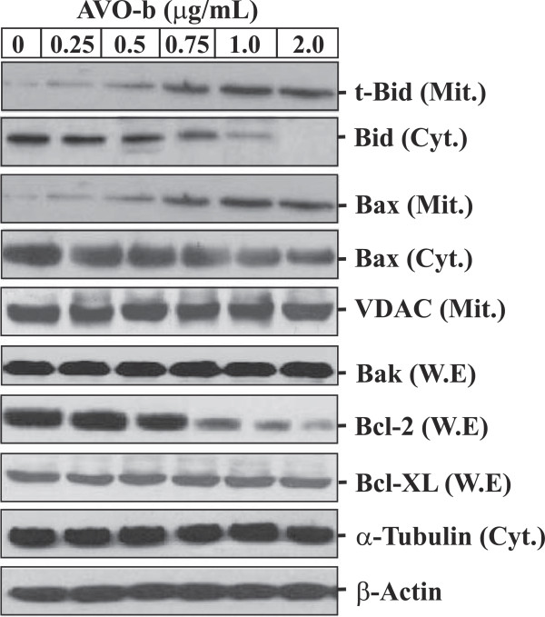
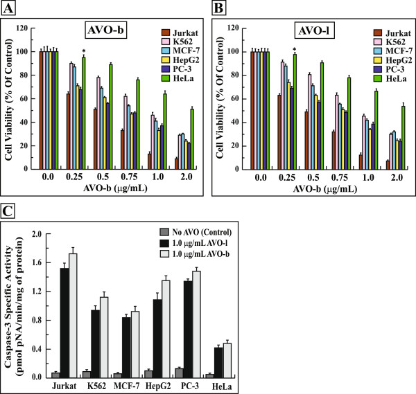
Results: Low concentrations of AVO-b and AVO-l inhibited the growth of HL-60 cells in a dose- and time-dependent manner. Employing flow cytometric, DNA fragmentation and caspase activation analyses, demonstrated that the cytotoxic effect of the oils is mediated by a caspase-dependent apoptosis. Kinetic studies in the presence and absence specific caspase inhibitors showed that activation of caspase-8 was dependent and subsequent to the activation of caspases-9 and -3. In addition, the essential oil caused a disruption of the mitochondrial transmembrane potential (ΔΨm), increased the release of cytochrome c to the cytosol, and altered the expression of certain members of Bcl-2 family (Bcl-2, Bax and Bid), Apaf-1 and XIAP. Interestingly, low doses of AVO-b and AVO-1 also induced apoptosis in various cancer cell lines, but not in noncancerous cells.

Conclusions: The results demonstrate that the EO-induced apoptosis in HL-60 cells is mediated by caspase-dependent pathways, involving caspases-3, -9, and -8, which are initiated by Bcl-2/Bax/Bid-dependent loss of ΔΨm leading to release of cytochrome c to the cytoplasm to activate the caspase cascade. The finding that AVO-b and AVO-l are more efficient to induce apoptosis in different cancer cell lines than noncancerous cells, suggests that A. vulgaris might be a promising source for new anticancer agents.


Figures

Figure 1
Figure 1
AVO inhibits the growth of HL-60 cells in a dose- and time-dependent manner. (A) HL-60 cells were treated with varying concentrations of AVO-l or AVO-b (0.0 to 2.0 μg/mL) for 24 h and the viability was analyzed by MTT assay as described in the methods. (B) HL-60 cells were incubated with 1.0 μg/mL of either AVO-l or AVO-b for different periods of time (0 to 48 h) and cell viability was assessed by the MTT assay. The control represents cells incubated under similar conditions in the absence of essential oil. The percentage of cell viability in each sample was expressed relative to untreated control, which was considered as a 100%. HL-60 cells were also treated with 8.0 μM etoposide under similar conditions as positive controls. The results shown represent the mean ± SD of three independent trials. Statistical analysis showed that all samples are significantly different (p < 0.05), when compared to untreated control without AVO or etoposide.
Figure 2
Figure 2
The growth inhibitory effect of AVO on HL-60 cells is mediated by apoptosis. (A) Cultured HL-60 cells were treated with varying concentration of AVO-b or AVO-l (0.0 to 2.0 μg/mL) an apoptosis was analyzed by a flow cytometer after staining with FITC-annexin-V and 7-AAD. The scattered blots showing the percentages of early and late apoptosis are indicated for one experiment. The graph represents the summary mean percentages of apoptosis (early and late apoptosis) of two independent experiments for each concentration of the oil. HL-60 cells were treated with 8.0 μM etoposide as a positive control. Statistical analysis showed that all samples are significantly different (p < 0.05), when compared to untreated controls. (B) Genomic DNA was extracted from HL-60 cells treated with the different concentrations of AVO-l or AVO-b, and applied to agarose gel electrophoresis. DNA fragmentation in the gel was visualized by UV light after staining with ethidium bromide. Mark, is a DNA ladder marker.
Figure 3
Figure 3
The apoptotic effect of AVO is mediated by activation of caspase cascades. (A) Whole cell extracts (50 μg), from HL-60 cells after treatment with varying concentrations of AVO-l or AVO-b for 24, were analyzed by Western blotting with antibodies against caspase-8 and -9 and caspase–3, respectively. The unprocessed forms of procaspase-8, -9 and -3, and their respective cleavage products of the active enzymes are indicated. The same membranes were also probed with an antibody against β-actin as a loading control. (B) Extracts (30 μg proteins) from cells treated with varying concentrations of AVO-l or AVO-b were analyzed for caspase-8, -9 and -3 catalytic activities using specific colorimetric tetrapeptide substrates. The specific enzyme activities, which represent the mean ± SD of three independent experiments, were measured as described in the methods. HL-60 cells were treated with 4.0 and 8.0 μM etoposide as positive controls. Statistical analysis showed that all samples are significantly different (p < 0.05), when compared to their untreated control. (C) HL-60 cells were incubated with either 35 μM of Caspase-8 inhibitor (z-IETD-fmk), caspase-9 inhibitor (z-LEHD-fmk), caspase-3 inhibitor (z-DEVD-fmk), or 50 μM of the general caspase inhibitor (z-VAD-fmk) for 6 h prior to addition of 1.0 μg/mL AVO-l or AVO-b for 24 h. After incubation, cell viability was assessed by the MTT assay. The cell viability for each sample treated with the specific caspase inhibitor was expressed as percentages relative to the parallel control cells incubated with the same caspase inhibitor alone, without the essential oil, which was considered 100%. The results shown represent the means ± SD of three independent experiments. Statistical analysis showed that all samples treated with caspase inhibitors and AVO are significantly different (p < 0.05), when compared to samples treated with the specific caspase inhibitor alone, AVO alone or untreated control.
Figure 4
Figure 4
AVO-b-induced processing and activation of caspase-8 in HL-60 cells requires activation of caspase-9 and -3. (A) HL-60 cells were treated with 1.0 μg/mL AVO-b for different time intervals (0 to 48 h), and caspase-9 (▲), caspase-8 (■) and caspase-3 (●) activities were determined calorimetrically and expressed as relative percentages to the control untreated cells. The data shown represent the means ± SD of two independent trials for each time point. (B) AVO-b-induced caspase-8 activation in the presence and absence (-) of 35 μM specific caspase-8 inhibitor (z-IETD-fmk), caspase-9 inhibitor (z-LEHD-fmk), caspase-3 inhibitor (z-DEVD-fmk), or 50 μM of the general caspase inhibitor (z-VAD-fmk). HL-60 cells were incubated with each of the individual caspase inhibitor for 6 hours before the exposure to 1.0 μg/mL AVO-b for an additional 24 h. Caspase-8 activation was analyzed calorimetrically after addition of the substrate Ac-IETD-pNA (the graph) and by immunoblotting with a specific anti-caspase-8 antibody (the Western blot). The results shown represent the percentages of caspase-8 activity relative to the control cells treated with AVO-b alone (-). “Cont.”, represents caspase-8 activity in HL-60 of untreated cells. Results of caspase-8 activity represent the means ± SD of three indecent trials. Statistical analysis showed that all samples treated with specific caspase inhibitors and AVO-b are significantly different (p < 0.05), when compared to cells treated with AVO-b alone.
Figure 5
Figure 5
AVO induces disruption of mitochondrial transmembrane potential (ΔΨm), promotes the release cytochrome c to the cytoplasm and modulates the expression of Apaf-1 and XIAP proteins in HL-60 cells. (A) HL-60 cells treated with 1.0 μg/ml AVO-l or AVO-b for different periods of time (0–24 h) and changes in mitochondrial membrane potential was monitored by a flourimetric analysis after addition of the fluorescent stain JC-1. Cells treated with 8.0 μM etoposide for 12 and 24 h were used as positive controls. The results shown represents the mean ± SD of three independent experiments. **Represents samples that are statistically different, when compared to their controls at 0 time or AVO untreated cells. (B) Western blot analysis of cytochrome c in the cytosolic fraction (50 μg) of HL-60 cells treated with AVO-b for different time points (0 to 48 h) as indicated at the top of each lane. (C) Western blot analysis of Apaf-1 and XIPA in whole cell extracts (W.E; 50 μg), and cytochrome c in cytosolic (Cyt.; 50 μg) and mitochondrial (Mit.; 30 μg) fractions obtained from HL-60 cells treated with varying concentrations of AVO-b (0.0–2.0 μg/mL). The same Western blots in both (B) and (C) for the cytosolic fractions and whole cell extracts were probed with antibodies to α-tubulin and β-actin, respectively, to ensure equal protein loading in the different lanes.
Figure 6
Figure 6
AVO-b modulates the expression of Bcl-2 family proteins, and promotes translocation of Bax and t-Bid to the mitochondria of HL-60 cells in a dose-dependent manner. Whole cell extracts (50 μg), mitochondrial (30 μg) and cytosolic (50 μg) fractions were prepared from HL-60 cells treated with AVO-b as described above. W.E were probed with antibodies against Bak, Bcl-2 and Bcl-xL proteins. Mit. fractions were immuneblotted with antibodies against t-Bid, Bax and VDAC, while Cyt. fractions were probed with antibodies for Bid and Bax. The same Western blots for the Cyt. fractions and W.E were probed with antibodies to α-tubulin and β-actin, respectively.
Figure 7
Figure 7
AVO inhibits the growth of various cancer cell lines by inducing a caspase-dependent apoptosis. Jurkat, K562, MCF-7, HepG2, PC-3 and HeLa cells were treated with varying concentrations (0.0-2.0 μg/mL) of AVO-b (A) or AVO-l (B) and the viability was analyzed by MTT assay. The data shown represent the mean ± SD of three independent experiments. (C) Extracts (30 μg proteins) from these cancer cells treated with 1.0 μg/mL of AVO-b or AVO-l were analyzed for caspase-3 catalytic activities. The specific enzyme activities, which represent the mean ± SD of three independent experiments, were measured as described in the methods. *p < 0.01 of statistically insignificant values, when compared to untreated samples.

Similar articles

Cited by

References

    1. Jemal A, Bray F, Center MM, Ferlay J, Ward E, Forman D. Global cancer statistics. CA Cancer J Clin. 2011;61(2):69–90. - PubMed
    1. Klein G. Cancer, apoptosis, and nonimmune surveillance. Cell Death Differ. 2004;11(1):13–17. - PubMed
    1. Hu W, Kavanagh JJ. Anticancer therapy targeting the apoptotic pathway. Lancet Oncol. 2003;4(12):721–729. - PubMed
    1. Kaufmann SH, Earnshaw WC. Induction of apoptosis by cancer chemotherapy. Exp Cell Res. 2000;256(1):42–49. - PubMed
    1. Alnemri ES. Mammalian cell death proteases: a family of highly conserved aspartate specific cysteine proteases. J Cell Biochem. 1997;64(1):33–42. - PubMed

Publication types

LinkOut - more resources