. 2014 Sep 1;127(Pt 17):3675-86.
doi: 10.1242/jcs.146282. Epub 2014 Jul 7.

PEX16 contributes to peroxisome maintenance by constantly trafficking PEX3 via the ER

Affiliations

PEX16 contributes to peroxisome maintenance by constantly trafficking PEX3 via the ER

Alexander Aranovich et al. J Cell Sci. .

Abstract

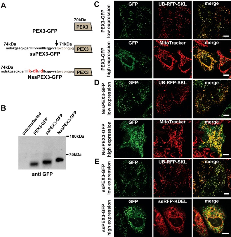
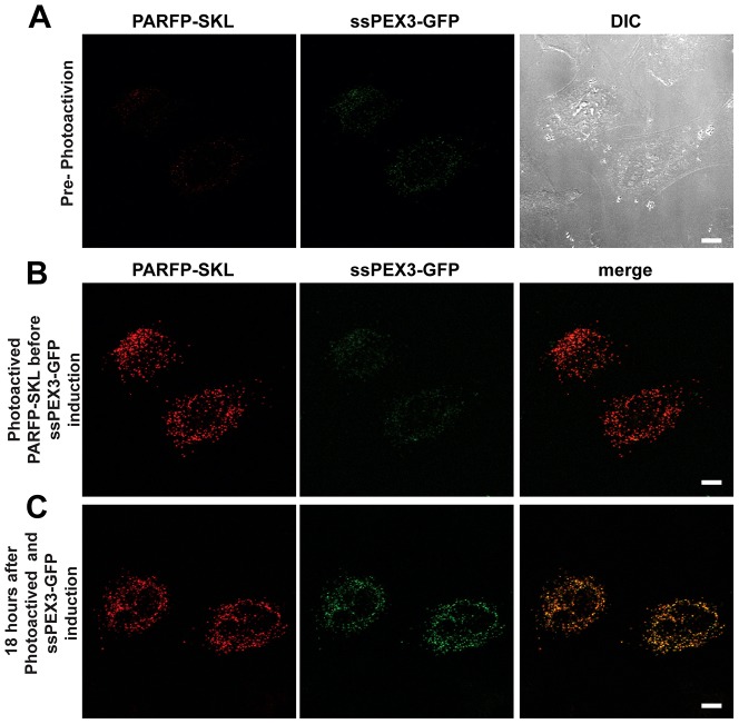
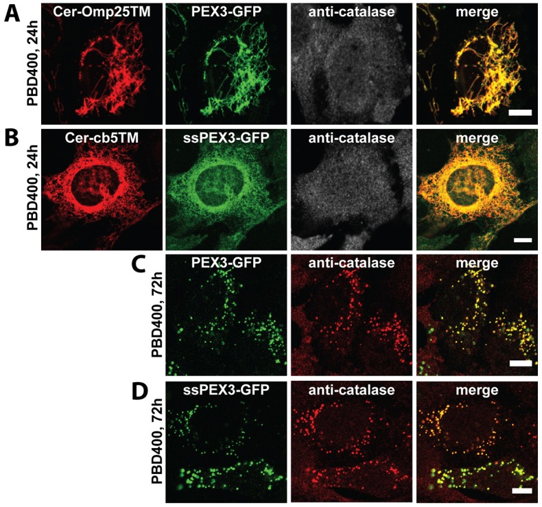
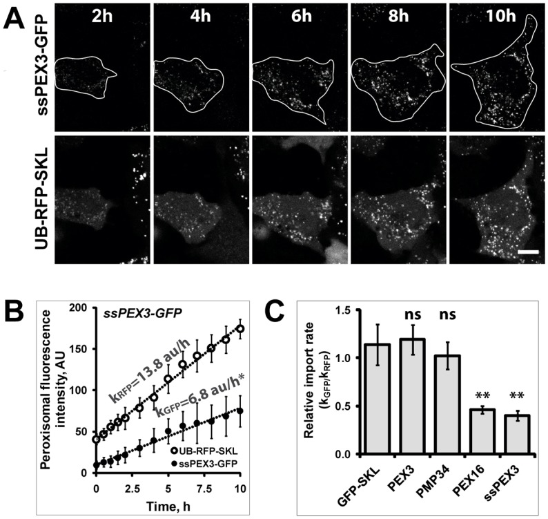
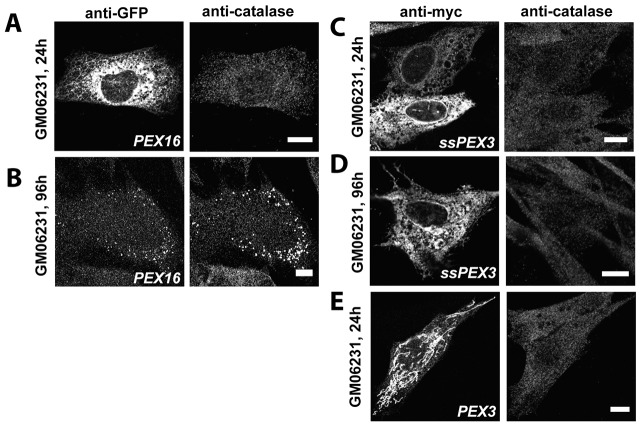
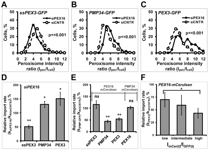
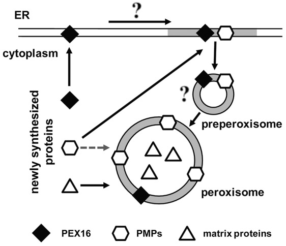
The endoplasmic reticulum (ER) is required for the de novo biogenesis of peroxisomes in mammalian cells. However, its role in peroxisome maintenance is unclear. To explore ER involvement in the maintenance of peroxisomes, we redirect a peroxisomal membrane protein (PMP), PEX3, to directly target to the ER using the N-terminal ER signal sequence from preprolactin. Using biochemical techniques and fluorescent imaging, we find that ER-targeting PEX3 (ssPEX3) is continuously imported into pre-existing peroxisomes. This suggests that the ER constitutively provides membrane proteins and associated lipids to pre-existing peroxisomes. Using quantitative time-lapse live-cell fluorescence microscopy applied to cells that were either depleted of or exogenously expressing PEX16, we find that PEX16 mediates the peroxisomal trafficking of two distinct peroxisomal membrane proteins, PEX3 and PMP34, via the ER. These results not only provide insight into peroxisome maintenance and PMP trafficking in mammalian cells but also highlight important similarities and differences in the mechanisms of PMP import between the mammalian and yeast systems.

Keywords: ER; Live cell imaging; Membrane trafficking; Organelle biogenesis; Peroxisomes; Protein trafficking.


Figures

Fig. 1.
Fig. 1.
ssPEX3-GFP protein is targeted to peroxisomes via the ER. (A) Representation of N-terminal ends of PEX3-GFP, ssPEX3-GFP and NssPEX3-GFP. GFP tagged at the C-terminus of each PEX3 construct is not shown. The amino acid sequences correspond to the signal sequence of preprolactin. The cleaved portion of the signal sequence is shown in bold, the arrow indicates the cleavage site. The calculated molecular mass of each construct, including the cleaved form of ssPEX3-GFP, is given in kDa. Mutations within the signal sequence that abolish the ER-targeting function of the signal sequence are highlighted in red for NssPEX3-GFP. (B) Signal sequence cleavage assay. Cells expressing PEX3-GFP, ssPEX3-GFP or NssPEX3-GFP were lysed 20 hours after transfection and analyzed by western blotting using an anti-GFP antibody. (C–E) Analysis of the subcellular localization of each PEX3 construct exogenously expressed in HeLa cells by using fluorescent live-cell microscopy 20 hours after transfection. Representative confocal fluorescence microscopy images for PEX3-GFP (C), NssPEX3-GFP (D) and ssPEX3-GFP (E) of both low and high expression levels are shown. For cells that expressed PEX3 at low levels, co-expression with the peroxisomal marker UB-RFP-SKL is shown. In images showing cells that express PEX3 at high levels, cells were stained with either MitoTracker Red (C,D) or they show co-expression of PEX3 with ER targeting ss-RFP-KDEL (E). See supplementary material Fig. S1 for magnification of high ssPEX3-GFP. All images were taken at the same settings and brightness for each panel was enhanced equally for presentation. Scale bars: 10 µm.
Fig. 2.
Fig. 2.
ssPEX3-GFP targets to pre-existing peroxisomes. HeLa Tet-On cells transfected with plasmids encoding ssPEX3-GFP under a TRE-Tight promoter and photoactivatable RFP-SKL (PARFP-SKL) under a CMV promoter. (A) Representative HeLa Tet-On cells before photoactivation of PARFP-SKL. Channels for PARFP-SKL, ssPEX3-GFP and DIC images are indicated. (B) Image of the same cell immediately after photoactivation of PARFP-SKL (red) followed by the addition of doxycycline to induce ssPEX3-GFP (green) expression (∼5 minutes). (C) The same cells as above 18 hours after photoactivation and induction. Scale bars: 10 µm.
Fig. 3.
Fig. 3.
ssPEX3-GFP and PEX3-GFP complement PEX3 deficiency in PBD400-T1 cells. Representative immunofluorescence confocal images of PEX3-deficient PBD400-T1 cells transiently co-transfected with plasmids encoding PEX3-GFP and mCerulean-Omp25TM (a mitochondrial marker) (A) or ssPEX3-GFP and mCerulean-cb5TM (an ER marker) (B) and immunostained for endogenous catalase 24 hours (A,B) and 72 hours after transfection (C,D), as indicated. The analyses of 300 cells expressing the PEX3 constructs 72 hours after transfection from three independent experiments resulted in almost 100% peroxisome recovery. Scale bars: 10 µm.
Fig. 4.
Fig. 4.
PMP import kinetic quantification assay. (A) Time-lapse imaging experiment of HeLa cells co-expressing ssPEX3-GFP with UB-RFP-SKL. GFP and RFP signals were acquired at 37°C over a period of 10 hours in CO2-independent medium in several different fields of view. Shown are representative time-lapse images at different time points. For presentation, the brightness of the images was enhanced equally for all frames. Scale bar: 10 µm. The boundaries of the analyzed cell are marked by white line on ssPEX3-GFP images. (B) The change in the fluorescent intensity within peroxisomes of ssPEX3-GFP (•) and UB-RFP-SKL (○) in the representative cell shown in A, is plotted against time to illustrate the rates of ssPEX3-GFP and UB-RFP-SKL import. The apparent peroxisome import kinetics is shown for both proteins. AU, arbitrary unit. Data were fit with linear regression (R2>0.98). Error bars indicate ±s.d. of signal between peroxisomes at a given time. Both the mean and the slopes were found to be significantly different (*P<0.001) by Student's t-test. (C) Summary for relative import rates of PEX3-GFP, PMP34-GFP, PEX16-GFP and ssPEX3-GFP (PEX3, PMP34, PEX16 and ssPEX3, respectively) respective to the import rate of UB-RFP-SKL (kGFP/kRFP) are shown in the bar graph. UB-GFP-SK (GFP-SKL) was used as a control. The analyses were performed on cells at early stages of protein expression. At least 17 cells from three independent experiments were analyzed for each GFP-protein construct. The error bars represent ±s.e.m. The P value was determined using Student's t-test. Both the relative import rates of PEX16 and ssPEX3 were significantly different from the GFP-SKL control (**P<0.005), whereas PEX3 and PMP34 showed no significant difference (ns). Scale bars: 10 µm.
Fig. 5.
Fig. 5.
Targeting of PEX3 to ER does not complement PEX16 deficient cells. Representative immunofluorescent confocal images of the PEX16-deficient cell line GM06231, transiently transfected with plasmids encoding PEX16-GFP (A,B), ssPEX3×3myc (C,D) or PEX3×3myc (E). PEX16-GFP was found initially in the ER (A) at 24 hours; however, at 96 hours it was found colocalized with catalase-positive punctate structures suggesting complementation of PEX16 function (B). In contrast, ssPEX3×3myc is unable to complement PEX16 deficiency in GM06231 cells (C,D). ssPEX3×3myc is localized to the ER at 24 (C) and 96 (D) hours after transfection. No catalase-positive punctuate structures were detected. (E) PEX3×3myc is localized to mitochondria in GM06231 cells. Scale bars: 10 µm.
Fig. 6.
Fig. 6.
Depletion of PEX16 expression delays ssPEX3-GFP import into peroxisome, whereas overexpression of PEX16 decreases the peroxisome import of PEX3-GFP and PMP34-GFP. (A–C) The distribution of PMP-GFP signal within peroxisomes (Iper.) with respect to the average fluorescence signal, within the whole cell (Icell) treated with either siPEX16 or siCNTR, are shown in a histogram as a ratio (Iper./Icell) for (A) ssPEX3-GFP, (B) PMP34-GFP, and (C) PEX3-GFP. The distributions for each PMP in siPEX16-treated cells were significantly different from that in control cells (siCNTR) (P<<0.001, n = 50). A scatter plot of the same data (A–C) is shown in supplementary material Fig. S3 (C–E). (D) The peroxisomal import rates [k(siPEX16)] of ssPEX3-GFP, PMP34-GFP, and PEX3-GFP in PEX16-depleted cells compared to siCNTR-treated [kAVsiCNTR)] cells are shown in a bar graph. (E) The peroxisomal import rates [k(PMP-GFP)] of ssPEX3-GFP, PMP34-GFP, PEX3-GFP or PEX16-GFP in cells co-expressing PEX16-mCerulean or PMP34-mCerulean, as indicated, compared to the average rates of the corresponding PMP-GFP construct in cells co-expressing mCerulean [kAV(CNTR)] are shown in a bar graph. (F) The rate of PEX3-GFP import into peroxisomes decreased with increasing PEX16-mCerulean expression. Shown here is a different representation of the data presented in E. Here, the relative import rate of PEX3-GFP compared to control is plotted based on its ratio of PEX16-mCerulean over the total PEX3-GFP fluorescence signal [ImCer(t)/IGFP(t)]. The rates are subdivided into three groups (with a comparable number of cells in each group) from low to high ImCer(t) to IGFP(t) ratio. The error bars indicate ±s.e.m. *P<0.05; **P<0.005; ns, not significant.
Fig. 7.
Fig. 7.
Model of PMP import into peroxisomes within mammalian cells. PMPs can be targeted to peroxisomes via two distinct pathways: the group I pathway through which PMPs initially target to the ER before being routed to peroxisomes; or the group II pathway through which PMPs are directly imported to peroxisomes. The pathway utilized by the PMP depends on the level of PEX16 in the ER, which is co-translationally targeted to the ER. On the ER, PEX16 can recruit other PMPs to the ER where they are rapidly transported to pre-existing peroxisomes. The mechanism of the transport between ER to peroxisomes has yet to be determined. However, based on studies using yeast and plant cells, PEX16 and PMPs may accumulate in a specialized domain on the ER that is enriched for PMPs and peroxisomal lipids (grey membrane) in order to generate pre-peroxisomal vesicles. Under conditions where PMPs are in excess compared to PEX16, such as an ectopic expression of PMPs, they can also target directly to peroxisomes via the group II pathway. Matrix proteins (triangles) are directly targeted to mature peroxisomes from the cytoplasm.

Similar articles

Cited by

References

    1. Brocard C. B., Boucher K. K., Jedeszko C., Kim P. K., Walton P. A. (2005). Requirement for microtubules and dynein motors in the earliest stages of peroxisome biogenesis. Traffic 6, 386–395 10.1111/j.1600-0854.2005.00283.x - DOI - PubMed
    1. Fang Y., Morrell J. C., Jones J. M., Gould S. J. (2004). PEX3 functions as a PEX19 docking factor in the import of class I peroxisomal membrane proteins. J. Cell Biol. 164, 863–875 10.1083/jcb.200311131 - DOI - PMC - PubMed
    1. Hoepfner D., Schildknegt D., Braakman I., Philippsen P., Tabak H. F. (2005). Contribution of the endoplasmic reticulum to peroxisome formation. Cell 122, 85–95 10.1016/j.cell.2005.04.025 - DOI - PubMed
    1. Honsho M., Tamura S., Shimozawa N., Suzuki Y., Kondo N., Fujiki Y. (1998). Mutation in PEX16 is causal in the peroxisome-deficient Zellweger syndrome of complementation group D. Am. J. Hum. Genet. 63, 1622–1630 10.1086/302161 - DOI - PMC - PubMed
    1. Honsho M., Hiroshige T., Fujiki Y. (2002). The membrane biogenesis peroxin Pex16p. Topogenesis and functional roles in peroxisomal membrane assembly. J. Biol. Chem. 277, 44513–44524 10.1074/jbc.M206139200 - DOI - PubMed