. 2014 Sep 1;127(Pt 17):3782-93.
doi: 10.1242/jcs.149849. Epub 2014 Jul 7.

Palmitoylation of plakophilin is required for desmosome assembly

Affiliations

Palmitoylation of plakophilin is required for desmosome assembly

Brett J Roberts et al. J Cell Sci. .

Abstract

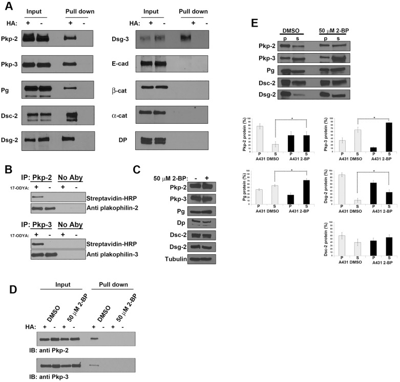
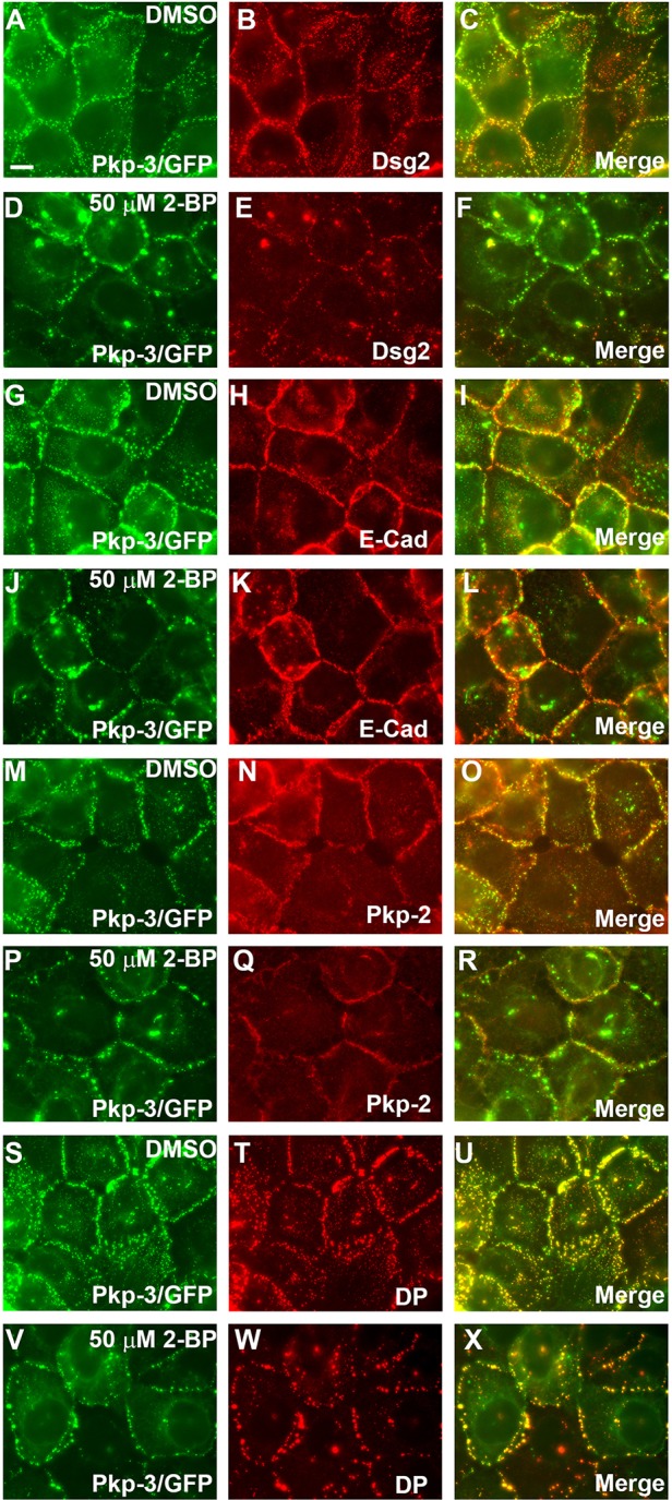
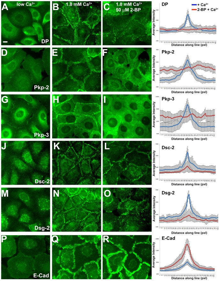
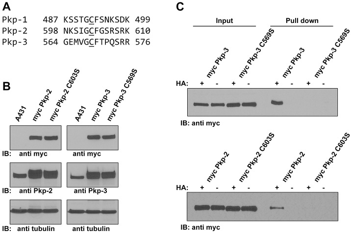
Desmosomes are prominent adhesive junctions found in various epithelial tissues. The cytoplasmic domains of desmosomal cadherins interact with a host of desmosomal plaque proteins, including plakophilins, plakoglobin and desmoplakin, which, in turn, recruit the intermediate filament cytoskeleton to sites of cell-cell contact. Although the individual components of the desmosome are known, mechanisms regulating the assembly of this junction are poorly understood. Protein palmitoylation is a posttranslational lipid modification that plays an important role in protein trafficking and function. Here, we demonstrate that multiple desmosomal components are palmitoylated in vivo. Pharmacologic inhibition of palmitoylation disrupts desmosome assembly at cell-cell borders. We mapped the site of plakophilin palmitoylation to a conserved cysteine residue present in the armadillo repeat domain. Mutation of this single cysteine residue prevents palmitoylation, disrupts plakophilin incorporation into the desmosomal plaque and prevents plakophilin-dependent desmosome assembly. Finally, plakophilin mutants unable to become palmitoylated act in a dominant-negative manner to disrupt proper localization of endogenous desmosome components and decrease desmosomal adhesion. Taken together, these data demonstrate that palmitoylation of desmosomal components is important for desmosome assembly and adhesion.

Keywords: Desmosome; Palmitoylation; Plakophilin.


Figures

Fig. 1.
Fig. 1.
Desmosomal components are palmitoylated. (A) A431 cell lysates were prepared, and ABE was performed to identify palmitoylated proteins. Lysates were split into two samples and processed with (+) and without (−) hydroxylamine (HA). After the final streptavidin–agarose pull-down, the captured proteins were resolved by SDS-PAGE, and individual desmosome and adherens junction components were detected by immunoblot analysis. Pkp, plakophilin; Pg, plakoglobin; DP, desmoplakin; Dsc-2, desmocollin-2; Dsg, desmoglein; E-cad, E-cadherin; α-cat, α-catenin; β-cat, β-catenin. (B) A431 cells were grown in medium with or without 100 µM 17-ODYA for 48 hours. Plakophilin-2 and plakophilin-3 were immunoprecipitated (IP) from cell lysates, and HRP–biotin was added to label cells using the Cu-catalyzed click reaction. Immunoprecipitations with no immunoprecipitating antibody (No Aby) served as negative controls. Immunoprecipitates were resolved by SDS-PAGE, and labeled proteins were detected by HRP–streptavidin or isoform-specific anti-plakophilin antibodies. (C) Cell lysates were prepared from A431 cells grown in the presence or absence of 50 µM 2-BP for 18 hours, and immunoblot analysis was performed to examine the levels of plakophilin-2, plakophilin-3, plakoglobin, desmoplakin, desmocollin-2 and desmoglein-2. β-tubulin was used as a loading control. (D) ABE demonstrated that plakophilin-2 and plakophilin-3 are palmitoylated in the absence of 2-BP, and palmitoylation is decreased in A431 cells grown in medium containing 50 µM 2-BP. IB, immunoblot. (E) Triton-X-100-insoluble (p) and -soluble (s) fractions were prepared from control A431 cultures (DMSO) or from A431 cells grown in medium containing 50 µM 2-BP. Immunoblot analysis determined that there was a significant increase in the soluble fraction of plakophilin-2, plakophilin-3, plakoglobin and desmoglein-2 in 2-BP-treated cultures. The Triton X-100 solubility of desmocollin-2 was unchanged following 2-BP treatment. Data show the mean±s.d. (n = 3 experiments); *P<0.05.
Fig. 2.
Fig. 2.
Inhibition of palmitoylation by 2-BP alters the localization of desmosomal components at cell borders. A431 cells expressing plakophilin-3–GFP (Pkp-3/GFP) were grown on glass coverslips in medium containing 50 µM 2-BP or DMSO as a negative control (18 hours). Cells were fixed and immunostained with antibodies specific for desmoglein-2 (Dsg2) (A–F), E-cadherin (E-Cad) (G–L), plakophilin-2 (Pkp-2) (M–R) and desmoplakin (DP) (S–X). Inhibition of palmitoylation has little effect on the localization of E-cadherin, whereas the localization of desmosomal components is disrupted at cell–cell borders. Scale bar: 10 µm.
Fig. 3.
Fig. 3.
Inhibition of palmitoylation disrupts Ca2+-induced desmosome assembly in HaCat keratinocytes. HaCat keratinocytes were grown on glass coverslips in medium containing low Ca2+ (left panels) in the absence (middle panels) or the presence of 50 µM 2-BP (right panels) overnight. Ca2+ was added (1.8 mM final concentration) to the medium for 2.5 hours and cells were processed for immunofluorescence microscopy using the antibodies indicated. Scale bar: 10 µm. Average fluorescence intensity was determined using Slidebook5 image software by measuring a segment of equal length and width across 30 individual cell borders for each treatment. Blue lines, the average fluorescence intensity for control cultures; red lines, fluorescence intensity in 2-BP-treated cell cultures. DP, desmoplakin; Pkp, plakophilin; Dsc-2, desmocollin-2; Dsg-2, desmoglein-2; E-cad, E-cadherin; Pxl, pixels. Data show the mean±s.d. (n = 30 cell border measurements).
Fig. 4.
Fig. 4.
A conserved cysteine in plakophilins is palmitoylated. (A) Sequence alignment of plakophilin-1 (NP_001005337), plakophilin-2 (NP_001005242) and plakophilin-3 (NP_009114) (Pkp-1, Pkp-2 and Pkp-3, respectively) shows similarity in the sequences surrounding the conserved cysteine residue. (B) Immunoblot (IB) analysis of cell lysates prepared from control A431 cells, A431 cells expressing Myc-tagged wild-type plakophilin-2 (myc Pkp-2), Myc-tagged plakophilin-2 C603S (myc Pkp-2 C603S), Myc-tagged wild-type plakophilin-3 (myc Pkp-3) and Myc-tagged plakophilin-3 C569S (myc Pkp-3 C569S). Lysates were blotted with antibodies against Myc, plakophilin-2 or plakophilin-3. Exogenous proteins were expressed at levels similar to those of the endogenous plakophilins, and the Myc-tagged proteins were all expressed at similar levels to one another. Tubulin is shown as a loading control. (C) ABE demonstrates that plakophilin-2 C603S and plakophilin-3 C569S are not palmitoylated, whereas wild-type plakophilin-2 and plakophilin-3 are palmitoylated. HA, hydroxylamine.
Fig. 5.
Fig. 5.
Palmitoylation-defective plakophilin-2 disrupts the localization of endogenous desmosomal components. A431 cells expressing wild-type plakophilin-2 (Pkp-2 WT; A–C, G–I and M–O) or plakophilin-2 C603S (Pkp-2 C603S; D–F, J–L and P–R) were grown on glass coverslips, fixed and immunostained using antibodies recognizing the Myc epitope tag (A,D), desmocollin-2 (Dsc-2; G,J) and desmoglein-2 (Dsg-2; M,P). Desmoplakin (DP) colocalization is shown in panels B, E, H, K, N and Q. Scale bar: 10 µm.
Fig. 6.
Fig. 6.
Plakophilin mutants display altered Triton X-100 solubility and lipid raft association compared with that of wild-type plakophilin. (A) Triton-X-100-insoluble (P) and soluble fractions (S) were prepared from A431 cells expressing wild-type plakophilin-2 (Pkp-2 WT), plakophilin-2 C603S (Pkp-2 C603S), wild-type plakophilin-3 (Pkp-3 WT) or plakophilin-3 C569S (Pkp-3 C569S). Immunoblot (IB) analysis was performed using antibodies against the Myc epitope tag, plakoglobin, desmoglein-2 and desmocollin-2/3. (B) Immunoblot analysis was performed, and the signal was quantified using a Li-Cor Odyssey Imaging system. Myc-tagged plakophilin-2 C603S and Myc-tagged plakophilin-3 C569S were found to be more soluble compared with Myc-tagged wild-type plakophilin-2 and plakophilin-3. The Triton X-100 solubility of endogenous plakoglobin (PG), desmoglein-2 (Dsg-2) and desmocollin-2/3 (Dsc-2) was not altered by the expression of mutant plakophilin-2 or plakophilin-3. (C,D) Cell lysates were prepared from A431 cells expressing Myc-tagged wild-type plakophilin-2 and Myc-tagged plakophilin-2 C603S (C) and Myc-tagged wild-type plakophilin-3 and Myc-tagged plakophilin-3 C569S (D). Lysates were subjected to sucrose-density centrifugation, 1-ml fractions were collected and immunoblot analysis was performed with the indicated antibodies. Fractions 4 and 5 (underlined) are enriched in the lipid raft component caveolin-1 (Cav-1), endogenous desmoglein-2 and wild-type plakophilin-2 and wild-type plakophilin-3. (E) The ratio of Myc signal to the desmoglein-2 signal present in fractions 4 and 5 is shown graphically. The ratios of plakophilin-2 C603S and plakophilin-3 C569S to the corresponding desmoglein-2 signal are reduced in fractions 4 and 5 compared with that of the wild-type plakophilins. For B,E, data show the mean±s.d. [n = 3 (B,E)]; *P<0.05.
Fig. 7.
Fig. 7.
Plakophilin-2 C603S fails to efficiently initiate desmosome assembly in A431DE cells. In the absence of 4OHT, neither plakophilin-2–ER (Pkp-2/ER) nor desmoplakin (DP) localize to cell borders (A–C). Addition of 100 nM 4OHT to the medium for 18 hours results in plakophilin-2–ER localization to cell borders and the recruitment of desmoplakin (D–F). In contrast to the wild-type protein, plakophilin-2-C603S–ER (Pkp-2 C603S/ER) did not efficiently localize to cell borders and desmoplakin was not efficiently recruited (G–I). Scale bar: 10 µm. (J) Immunoblot (IB) analysis of A431DE cells expressing plakophilin-2–ER or plakophilin-2-C603S–ER demonstrates equivalent expression of the plakophilin-2 fusion proteins. β-tubulin is included as a loading control (K) A431DE cells expressing plakophilin-2–ER or plakophilin-2-C603S–ER were grown in medium containing 100 nM 4OHT for 18 hours. Triton-X-100-soluble (S) and Triton-X-100-insoluble pellet (P) fractions were prepared and immunoblotted with antibodies against plakophilin-2. Data were collected using Li-Cor Odyssey near-infrared imaging. Data show the mean±s.d. (n = 3); **P<0.01.
Fig. 8.
Fig. 8.
Expression of plakophilin palmitoylation mutants disrupts desmosomal adhesion. Dispase adhesion assays were performed to examine the relative strength of desmosomal adhesion in parental A431 cells (A) and in A431 cells expressing Myc-tagged plakophilin-2 (Pkp-2; B), Myc-tagged plakophilin-2 C603S (C), Myc-tagged plakophilin-3 (Pkp-3; D) and Myc-tagged plakophilin-3 C569S (E). A–E depict representative examples of cell sheet fragments counted in the dispase assay. (F) Quantification of the dispase assays. Data show the mean±s.d. (n = 3); *P<0.05.

Similar articles

Cited by

References

    1. Aicart-Ramos C., Valero R. A., Rodriguez-Crespo I. (2011). Protein palmitoylation and subcellular trafficking. Biochim. Biophys. Acta 1808, 2981–2994 10.1016/j.bbamem.2011.07.009 - DOI - PubMed
    1. Bazzi H., Getz A., Mahoney M. G., Ishida-Yamamoto A., Langbein L., Wahl J. K., 3rd, Christiano A. M. (2006). Desmoglein 4 is expressed in highly differentiated keratinocytes and trichocytes in human epidermis and hair follicle. Differentiation 74, 129–140 10.1111/j.1432-0436.2006.00061.x - DOI - PubMed
    1. Bonné S., Gilbert B., Hatzfeld M., Chen X., Green K. J., van Roy F. (2003). Defining desmosomal plakophilin-3 interactions. J. Cell Biol. 161, 403–416 10.1083/jcb.200303036 - DOI - PMC - PubMed
    1. Boukamp P., Petrussevska R. T., Breitkreutz D., Hornung J., Markham A., Fusenig N. E. (1988). Normal keratinization in a spontaneously immortalized aneuploid human keratinocyte cell line. J. Cell Biol. 106, 761–771 10.1083/jcb.106.3.761 - DOI - PMC - PubMed
    1. Brennan D., Peltonen S., Dowling A., Medhat W., Green K. J., Wahl J. K., 3rd, Del Galdo F., Mahoney M. G. (2012). A role for caveolin-1 in desmoglein binding and desmosome dynamics. Oncogene 31, 1636–1648 10.1038/onc.2011.346 - DOI - PMC - PubMed

Publication types