. 2014 Sep;80(18):5698-708.
doi: 10.1128/AEM.01196-14. Epub 2014 Jul 7.

Chitin accelerates activation of a novel haloarchaeal serine protease that deproteinizes chitin-containing biomass

Affiliations

Chitin accelerates activation of a novel haloarchaeal serine protease that deproteinizes chitin-containing biomass

Yaoxin Zhang et al. Appl Environ Microbiol. 2014 Sep.

Abstract

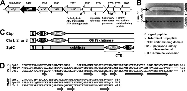
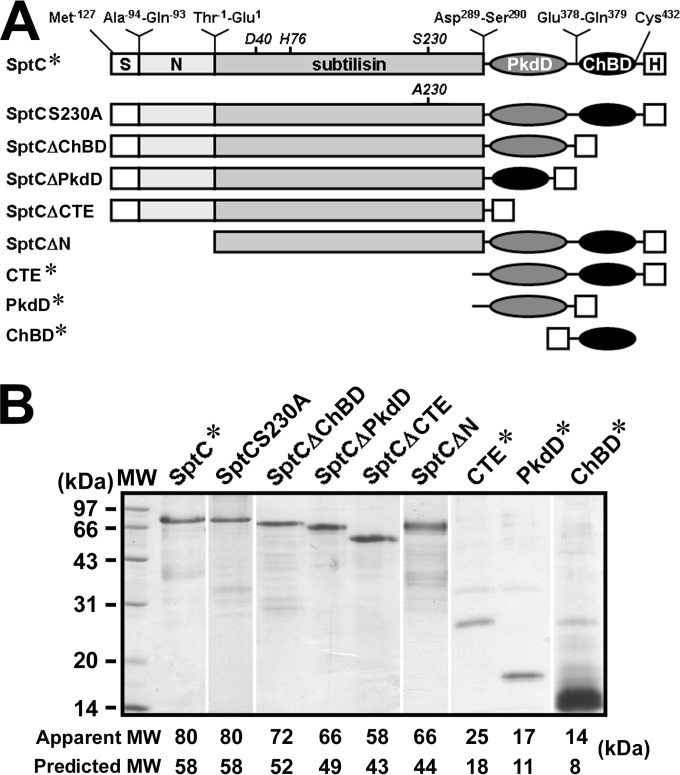
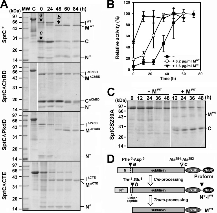
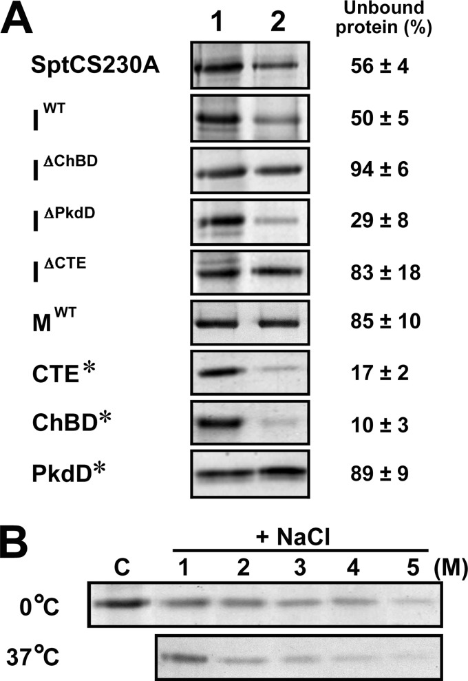
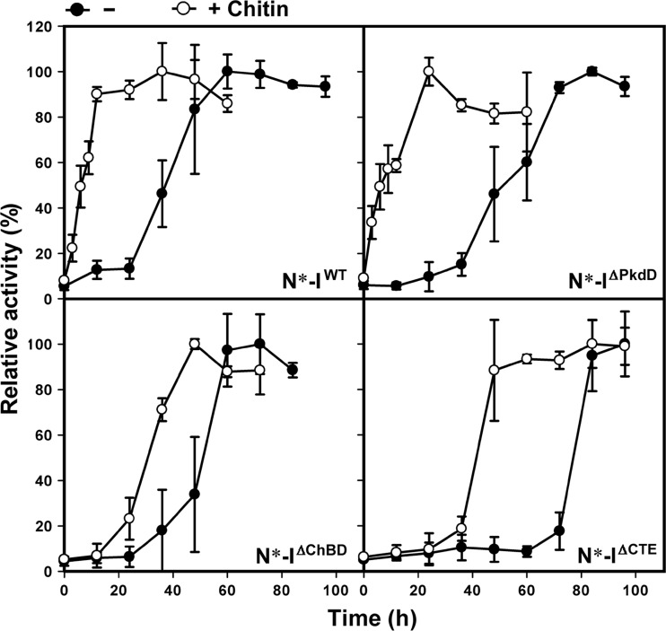
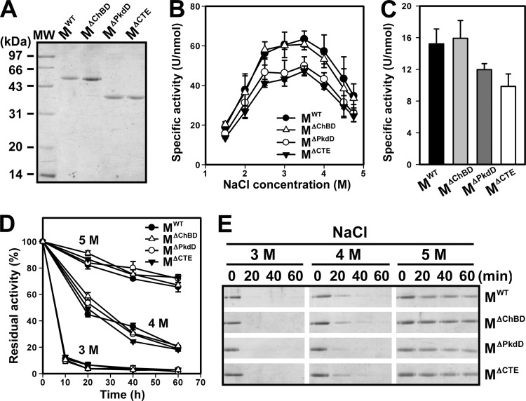
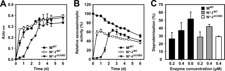
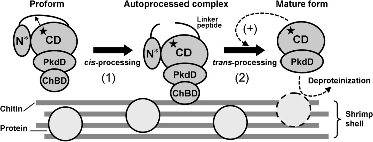
The haloarchaeon Natrinema sp. strain J7-2 has the ability to degrade chitin, and its genome harbors a chitin metabolism-related gene cluster that contains a halolysin gene, sptC. The sptC gene encodes a precursor composed of a signal peptide, an N-terminal propeptide consisting of a core domain (N*) and a linker peptide, a subtilisin-like catalytic domain, a polycystic kidney disease domain (PkdD), and a chitin-binding domain (ChBD). Here we report that the autocatalytic maturation of SptC is initiated by cis-processing of N* to yield an autoprocessed complex (N*-I(WT)), followed by trans-processing/degradation of the linker peptide, the ChBD, and N*. The resulting mature form (M(WT)) containing the catalytic domain and the PkdD showed optimum azocaseinolytic activity at 3 to 3.5 M NaCl, demonstrating salt-dependent stability. Deletion analysis revealed that the PkdD did not confer extra stability on the enzyme but did contribute to enzymatic activity. The ChBD exhibited salt-dependent chitin-binding capacity and mediated the binding of N*-I(WT) to chitin. ChBD-mediated chitin binding enhances SptC maturation by promoting activation of the autoprocessed complex. Our results also demonstrate that SptC is capable of removing proteins from shrimp shell powder (SSP) at high salt concentrations. Interestingly, N*-I(WT) released soluble peptides from SSP faster than did M(WT). Most likely, ChBD-mediated binding of the autoprocessed complex to chitin in SSP not only accelerates enzyme activation but also facilitates the deproteinization process by increasing the local protease concentration around the substrate. By virtue of these properties, SptC is highly attractive for use in preparation of chitin from chitin-containing biomass.


Figures

FIG 1
FIG 1
Organization of the chitin metabolism-related gene cluster in Natrinema sp. J7-2 and the primary structural features of SptC. (A) Schematic illustrating the organization of the chitin metabolism-related gene cluster in strain J7-2. Genes encoding chitin-binding protein (Cbp), SptC, chitinases (Chi1, Chi2, and Chi3), and proteins involved in carbohydrate transport and metabolism (NJ7G_2703-2707) are drawn to scale as arrows. The locus tag of each gene is shown above the corresponding arrow. (B) Assay of chitinolytic activity of strain J7-2. Strain J7-2 was cultured at 37°C for 2 weeks on plates containing basal medium with (+) or without (−) 1 g/liter of colloidal chitin. (C) Schematic representation of the domain organization in Cbp, Chi1, Chi2, Chi3, and SptC. (D) Amino acid sequence alignments of the PkdD and ChBD in SptC (GenBank accession number AFO57926) with those of chitin-binding protease AprIV (GenBank accession number BAB86297) from P. piscicida strain O-7, the PkdD (3JQU) of collagenase (ColG) from C. histolyticum, and the ChBD (1E15) of chitinase B (ChiB) from S. marcescens. The amino acid residues of SptC are numbered starting from the N terminus of the precursor. The β-strands in the PkdD of ColG and the ChBD of ChiB are indicated by horizontal arrows.
FIG 2
FIG 2
Schematic representation of the primary structures (A) and SDS-PAGE analysis of recombinant SptC and its derivatives (B). (A) The locations of the active-site residues (D40, H76, and S230) in the subtilisin-like catalytic domain and the N- and C-terminal residues of each region are shown. S, N, and H represent the signal peptide, N-terminal propeptide, and His tag, respectively. (B) Samples of purified proteins were precipitated with TCA and then subjected to SDS-PAGE analysis. The predicted molecular mass (MW) of each protein was calculated based on the amino acid sequence, and the apparent MW was determined by SDS-PAGE analysis.
FIG 3
FIG 3
Autoprocessing of recombinant SptC and its derivatives. (A) SDS-PAGE analysis of the autoprocessing of SptC* and its derivatives. Samples of purified SptC* and its derivatives (5 μg/ml, lane C) in buffer A (50 mM Tris-HCl, 10 mM CaCl2 [pH 8.0]) containing 8 M urea and 10 mM β-ME were dialyzed overnight at 4°C against the same buffer containing 5 M NaCl (lane 0) and then incubated at 40°C. At the indicated times, aliquots were withdrawn and subjected to SDS-PAGE analysis. The bands labeled a, b, and c were subjected to N-terminal sequencing. (B) Activation of the autoprocessed complex. N*-IWT (5 μg/ml) was incubated at 40°C in buffer A containing 5 M NaCl in the absence (−) or presence (+) of 0.2 or 1.6 μg/ml of MWT. At the indicated times, aliquots were removed and subjected to azocaseinolytic-activity assay. Values are expressed as the means ± SDs (bars) of three independent experiments, and the highest mean activity value observed during the maturation process was defined as 100% to determine relative activity. (C) Processing of SptCS230A by mature SptC. SptCS230A (5 μg/ml, lane 0) in buffer A containing 5 M NaCl was incubated at 40°C in the absence (−) or presence (+) of 0.3 μg/ml of MWT. At the indicated times, aliquots were removed and subjected to SDS-PAGE analysis. (D) Schematic representation of the autoprocessing of the proform of SptC. Filled arrowheads indicate the autoprocessing sites, and the open arrowhead indicates the site of proteolytic cleavage of SptC by the active mature enzyme. The identified N termini of bands a, b, and c from panel A are indicated. The positions of the intermediates (IWT, IΔChBD, IΔPkdD, and IΔCTE), the mature forms (MWT, MΔChBD, MΔPkdD, and MΔCTE), the processed core domain of the N-terminal propeptide (N*), and the cleaved C-terminal fragments (C and CΔChBD) are also indicated to the right of each gel in panels A and C.
FIG 4
FIG 4
ChBD-mediated binding of SptC to chitin. (A) Chitin-binding capacities of SptCS230A, the intermediate forms (IWT, IΔChBD, IΔPkdD, and IΔCTE), the mature form (MWT), and the C-terminal fragments (ChBD*, PkdD*, and CTE*). Each protein (5 μg/ml) was incubated in 300 μl of buffer A (50 mM Tris-HCl, 10 mM CaCl2 [pH 8.0]) containing 5 M NaCl at 0°C for 30 min in the absence (lane 1) or presence (lane 2) of 5 mg of insoluble chitin. After centrifugation, 120 μl of the supernatant was subjected to SDS-PAGE analysis. Numbers given on the right of the images indicate the densitometric ratios of unbound protein (lane 2) to the control (lane 1) and are expressed as the means ± SDs of three independent experiments. (B) Salt dependence of the binding of the CTE* to chitin. CTE* (5 μg/ml) was incubated at 0 or 37°C for 30 min without chitin and NaCl (lane C) or with 5 mg of insoluble chitin in 300 μl of buffer A containing different concentrations of NaCl (1 to 5 M). After centrifugation, the supernatants were subjected to SDS-PAGE analysis.
FIG 5
FIG 5
Chitin-accelerated activation of SptC. Autoprocessed complex (N*-IWT, N*-IΔChBD, N*-IΔPkdD, or N*-IΔCTE) at a concentration of 5 μg/ml was kept standing and incubated 40°C in buffer A (50 mM Tris-HCl, 10 mM CaCl2 [pH 8.0]) containing 5 M NaCl at in the absence (−) or presence (+) of 4 mg/ml of chitin. At the indicated times, aliquots were removed and subjected to azocaseinolytic-activity assay. Values are expressed as the means ± SDs (bars) of three independent experiments, and the highest mean activity value observed during the maturation process was defined as 100% to determine relative activity.
FIG 6
FIG 6
Properties of the mature enzymes. (A) SDS-PAGE analysis of the purified mature enzymes. (B) Salt dependence of enzyme activity. The azocaseinolytic activity of the mature enzymes was measured at 40°C in buffer A containing different concentrations of NaCl. (C) Enzymatic hydrolysis of suc-AAPF-pNA. Enzyme activity was measured at 40°C using suc-AAPF-pNA (0.1 mM) as the substrate in buffer A containing 3.5 M NaCl. (D and E) Heat inactivation of the mature enzymes. The mature enzymes (3 μg/ml) were incubated at 60°C in buffer A (50 mM Tris-HCl, 10 mM CaCl2 [pH 8.0]) containing 3, 4, or 5 M NaCl. At the indicated times, aliquots were removed and subjected to azocaseinolytic-activity assay (D) and SDS-PAGE analysis (E). Residual activity is expressed as a percentage of the original activity of each sample (D). Values (B, C, and D) are expressed as the means ± SDs (bars) of three independent experiments.
FIG 7
FIG 7
Deproteinization of SSP by SptC. (A and B) Release of soluble peptides from SSP by enzymatic treatment (A) and proteolytic-activity assay of the reaction mixture (B). SSP (2 mg) was incubated with MWT, N*-IWT, or N*-IΔChBD (0.4 μM) at 40°C in 200 μl of buffer A (50 mM Tris-HCl, 10 mM CaCl2 [pH 8.0]) containing 5 M NaCl. At the indicated times, the soluble fraction was recovered from the reaction mixture by centrifugation and subjected to absorbance measurement at 280 nm (A) and azocaseinolytic-activity assay (B). Relative azocaseinolytic activity was calculated with the original activity of the MWT-treated sample, defined as 100%. (C) Percent deproteinization of SSP using different enzyme samples. After 6 days of treatment with different concentrations of MWT (0.2 to 0.6 μM), N*-IWT (0.2 to 0.4 μM), or 0.4 μM N*-IΔChBD as described above, SSP in the reaction mixture was recovered by centrifugation and the degree of deproteinization was determined. For reference purposes, 0% deproteinization was defined as 24.4 mg of protein contained in 1 g of SSP without enzyme treatment. Values are expressed as the means ± SDs (bars) of three independent experiments.
FIG 8
FIG 8
Proposed model for autocatalytic maturation of SptC. The core domain (N*) of the N-terminal propeptide in the proform is cis-processed by the active site of the catalytic domain (CD) to yield the autoprocessed complex (step 1). The active site is indicated by a star. Next, the linker peptide and the ChBD are trans-processed and N* is degraded, generating the active mature form containing the CD and the PkdD (step 2). Active enzyme that matured earlier can catalyze (+) the step 2 reaction to accelerate the maturation process. ChBD-mediated binding of the autoprocessed complex to chitin not only accelerates conversion of the complex into the mature form but also increases the local enzyme concentration to promote enzyme activation and improve the deproteinization of chitin-containing biomass, such as shrimp shell (see Discussion for details). Note that minerals, lipids, and pigments in chitin-containing biomass are not shown.

Similar articles

Cited by

References

    1. De Castro RE, Maupin-Furlow JA, Gimenez MI, Herrera Seitz MK, Sanchez JJ. 2006. Haloarchaeal proteases and proteolytic systems. FEMS Microbiol. Rev. 30:17–35. 10.1111/j.1574-6976.2005.00003.x - DOI - PubMed
    1. Siezen RJ, Leunissen JAM. 1997. Subtilases: the superfamily of subtilisin-like serine proteases. Protein Sci. 6:501–523. 10.1002/pro.5560060301 - DOI - PMC - PubMed
    1. Kamekura M, Seno Y, Holmes ML, Dyall-Smith ML. 1992. Molecular cloning and sequencing of the gene for a halophilic alkaline serine protease (halolysin) from an unidentified halophilic archaea strain (172P1) and expression of the gene in Haloferax volcanii. J. Bacteriol. 174:736–742 - PMC - PubMed
    1. Ryu K, Kim J, Dordick JS. 1994. Catalytic properties and potential of an extracellular protease from an extreme halophile. Enzyme Microb. Technol. 16:266–275. 10.1016/0141-0229(94)90165-1 - DOI - PubMed
    1. Ruiz DM, Iannuci NB, Cascone O, De Castro RE. 2010. Peptide synthesis catalysed by a haloalkaliphilic serine protease from the archaeon Natrialba magadii (Nep). Lett. Appl. Microbiol. 51:691–696. 10.1111/j.1472-765X.2010.02955.x - DOI - PubMed

Publication types

MeSH terms

LinkOut - more resources