. 2014 Jul 22;111(29):E2937-46.
doi: 10.1073/pnas.1406381111. Epub 2014 Jul 7.

Molecular mechanisms for the regulation of histone mRNA stem-loop-binding protein by phosphorylation

Affiliations

Molecular mechanisms for the regulation of histone mRNA stem-loop-binding protein by phosphorylation

Jun Zhang et al. Proc Natl Acad Sci U S A. .

Abstract

Replication-dependent histone mRNAs end with a conserved stem loop that is recognized by stem-loop-binding protein (SLBP). The minimal RNA-processing domain of SLBP is phosphorylated at an internal threonine, and Drosophila SLBP (dSLBP) also is phosphorylated at four serines in its 18-aa C-terminal tail. We show that phosphorylation of dSLBP increases RNA-binding affinity dramatically, and we use structural and biophysical analyses of dSLBP and a crystal structure of human SLBP phosphorylated on the internal threonine to understand the striking improvement in RNA binding. Together these results suggest that, although the C-terminal tail of dSLBP does not contact the RNA, phosphorylation of the tail promotes SLBP conformations competent for RNA binding and thereby appears to reduce the entropic penalty for the association. Increased negative charge in this C-terminal tail balances positively charged residues, allowing a more compact ensemble of structures in the absence of RNA.

Keywords: NMR; X-ray crystallography; intrinsically disordered protein.


Conflict of interest statement

The authors declare no conflict of interest.

Figures

Fig. 1.
Fig. 1.
Schematic of the domain architecture of dSLBP (Upper) and amino acid sequence alignment of RPDs of Drosophila and human SLBP (Lower). Domains of SLBP include the N-terminal domain (NTD), RBD, and C-terminal region (C). Amino acid sequences are shown with the RBD sequence in the top two rows and the C-terminal region in the bottom row. T230 in the TPNK motif and phosphorylation sites in the C-terminal region are indicated with boldface and asterisks, respectively; the residues involved in RNA binding are shown in cyan; and acidic residues in the C-terminal region are shown in red.
Fig. 2.
Fig. 2.
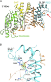
Crystal structure of the phosphorylated hSLBP RBD-pT171 in complex with the histone mRNA stem loop and 3′hExo. (A) Superposition of the crystal structure of the human histone mRNA stem loop (orange), hSLBP RBD-pT171 (cyan), and 3′hExo (green nuclease domain and yellow SAP domain) ternary complex with that of the unphosphorylated ternary complex (gray) (18). The red arrow points to the loop region that is disordered in the unphosphorylated complex. (B) Interaction of hSLBP side chains with pT171. Hydrogen bonds and salt bridges involving the phosphate group on T171 are shown by red dashes. The red sphere is a water molecule that interacts with both W190 and the phosphate group. Simulated annealing omit Fobs–Fcalc electron density for the phosphate is shown also, contoured at 3σ. Equivalent residues in hSLBP/dSLBP are as follows: T171/T230, K146/K206, Y151/Y211, R160/K220, R163/R223, and W190/W249. This figure, Figs. 3 and 6F, and Figs. S3 and S6 were created with PyMOL (Schrödinger).
Fig. 3.
Fig. 3.
Crystal structures of dSLBP RPD in complex with the histone mRNA stem loop. (A) Ribbon diagram of dSLBP RPD-5E in complex with the histone mRNA stem loop. A disordered region in the αB–αC loop (K220–T224) is indicated by small spheres. (B) Superposition of the crystal structures of protein:RNA complexes of dSLBP RPD-5E (aqua), dSLBP RPD-WT (green), and hSLBP RPD. (PDB ID code 4HXH is shown in cyan).
Fig. 4.
Fig. 4.
NMR analysis of the dSLBP RPD-5E:RNA complex. (A) 15N-HSQC of free (red) and RNA-bound (black) dSLBP RPD-5E. For clarity, only the assignments for the residues involved in RNA binding are labeled. (B) Methyl regions of the 1D-1H spectrum of free (red) and RNA-bound (black) dSLBP RPD-5E. Peaks for methyl groups shifted to high field are starred. (C) Plot of relative peak intensities of 15N-HSQC of RNA-bound dSLBP RPD-5E, as shown in A. The C-terminal region (C-term) is denoted by the purple bar. The peak intensities of unassigned residues are not shown and appear as gaps in the histogram.
Fig. 5.
Fig. 5.
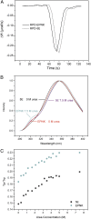
ITC and FRET studies of dSLBP RPD. (A) ITC measurements of histone mRNA stem-loop binding for dSLBP RPD-EPNK and dSLBP RPD-5E. Representative curves are shown. (B) Representative fluorescence emission curves of free dSLBP RPD-5E and dSLBP RPD-EPNK at 0 and 7.5 M urea. Curves for dSLBP RPD-5E are shown in black (0 M urea) and purple (7.5 M urea), and curves for dSLBP RPD-EPNK are shown in aqua (0 M urea) and red (7.5 M urea). The curves are normalized to the emission of W249. (C) Ratio of Tyr/Trp emission with increasing urea concentration for free dSLBP RPD-5E and dSLBP RPD-EPNK.
Fig. 6.
Fig. 6.
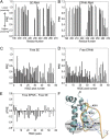
PRE study of dSLBP RPD. PRE ratios are plotted for RNA-bound and free dSLBP RPD: (A) RNA-bound RPD-5E, (B) RNA-bound RPD-EPNK, (C) free RPD-5E, and (D) free RPD-EPNK. PRE values for unassigned residues in A and B are not shown. Resonances were partially assigned for C and D; the assigned peaks are shown in black and the unassigned peaks are shown in gray. (E) Plot of differences between PRE ratios for free dSLBP RPD-EPNK and dSLBP RPD-5E. (F) Location of residues with positive PRE ratio differences, as shown in E. A ribbon diagram of dSLBP RPD-5E in complex with histone mRNA stem loop is shown displaying side chains for a Ser-Arg-Arg motif, S196, R197, and R198 (gray oval), whose assigned resonances have positive ΔPRE ratios. Side chains for a second Ser-Arg-Arg motif (gray oval) and additional basic residues on the RNA-binding surface are shown also.
Fig. 7.
Fig. 7.
Model for phosphorylation-stimulated binding of dSLBP to the histone mRNA stem loop. Nonphosphorylated (WT) dSLBP exists as an ensemble of conformations (Left) that becomes more compact upon phosphorylation (indicated by purple spheres) of the TPNK motif and C-terminal tail (Center). The phosphorylated C-terminal tail is flexible upon RNA binding and available to bind other molecules (Right).

Similar articles

Cited by

References

    1. Harris ME, et al. Regulation of histone mRNA in the unperturbed cell cycle: Evidence suggesting control at two posttranscriptional steps. Mol Cell Biol. 1991;11(5):2416–2424. - PMC - PubMed
    1. Marzluff WF, Wagner EJ, Duronio RJ. Metabolism and regulation of canonical histone mRNAs: Life without a poly(A) tail. Nat Rev Genet. 2008;9(11):843–854. - PMC - PubMed
    1. Marzluff WF, Duronio RJ. Histone mRNA expression: Multiple levels of cell cycle regulation and important developmental consequences. Curr Opin Cell Biol. 2002;14(6):692–699. - PubMed
    1. Dominski Z, Marzluff WF. Formation of the 3′ end of histone mRNA. Gene. 1999;239(1):1–14. - PubMed
    1. Birnstiel ML, Busslinger M, Strub K. Transcription termination and 3′ processing: The end is in site! Cell. 1985;41(2):349–359. - PubMed

Publication types

MeSH terms

LinkOut - more resources