. 2014 Aug 29;289(35):24263-74.
doi: 10.1074/jbc.M114.589408. Epub 2014 Jul 7.

Structural basis for regulation of the human acetyl-CoA thioesterase 12 and interactions with the steroidogenic acute regulatory protein-related lipid transfer (START) domain

Affiliations

Structural basis for regulation of the human acetyl-CoA thioesterase 12 and interactions with the steroidogenic acute regulatory protein-related lipid transfer (START) domain

Crystall M D Swarbrick et al. J Biol Chem. .

Abstract

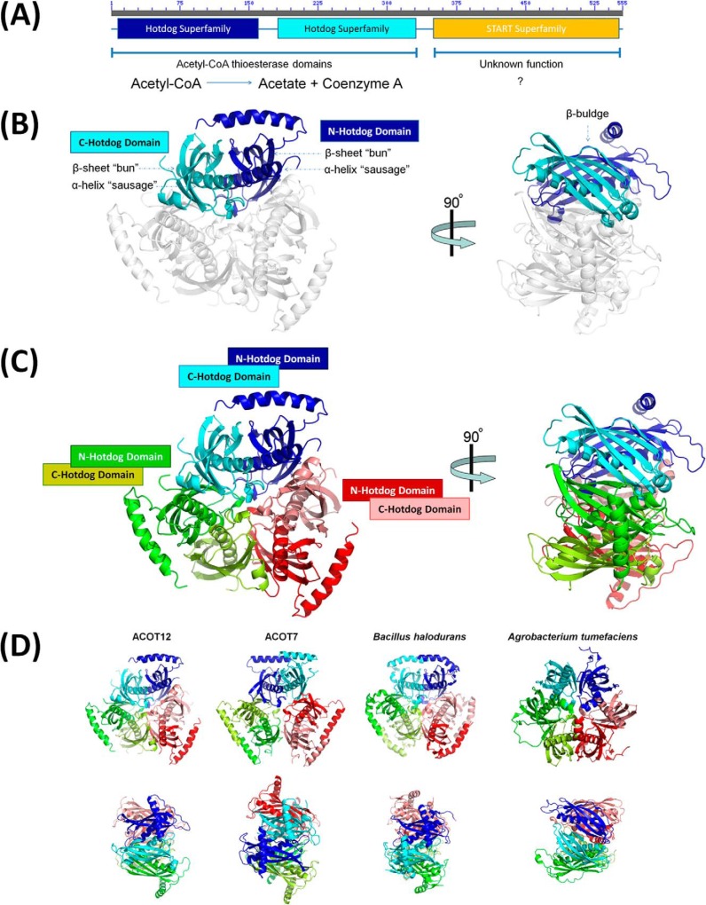
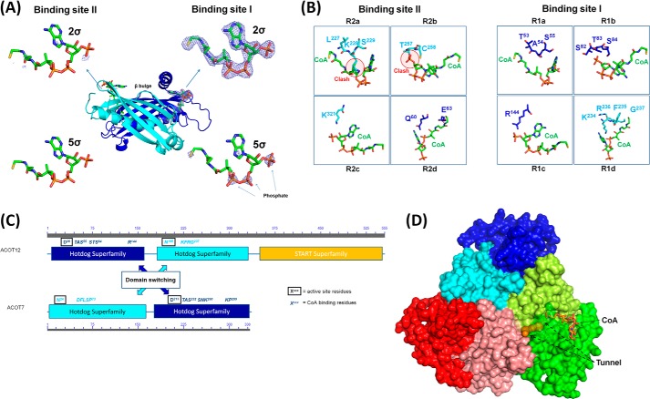
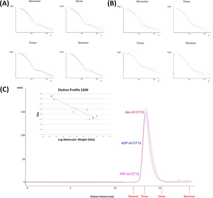
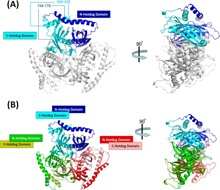
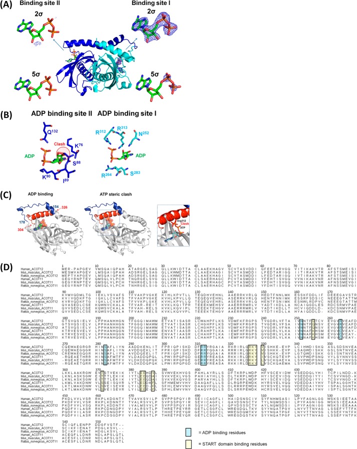
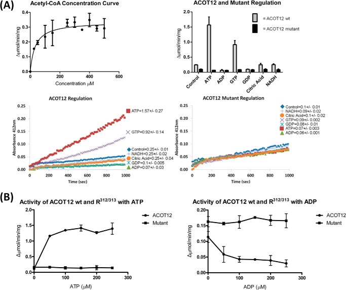
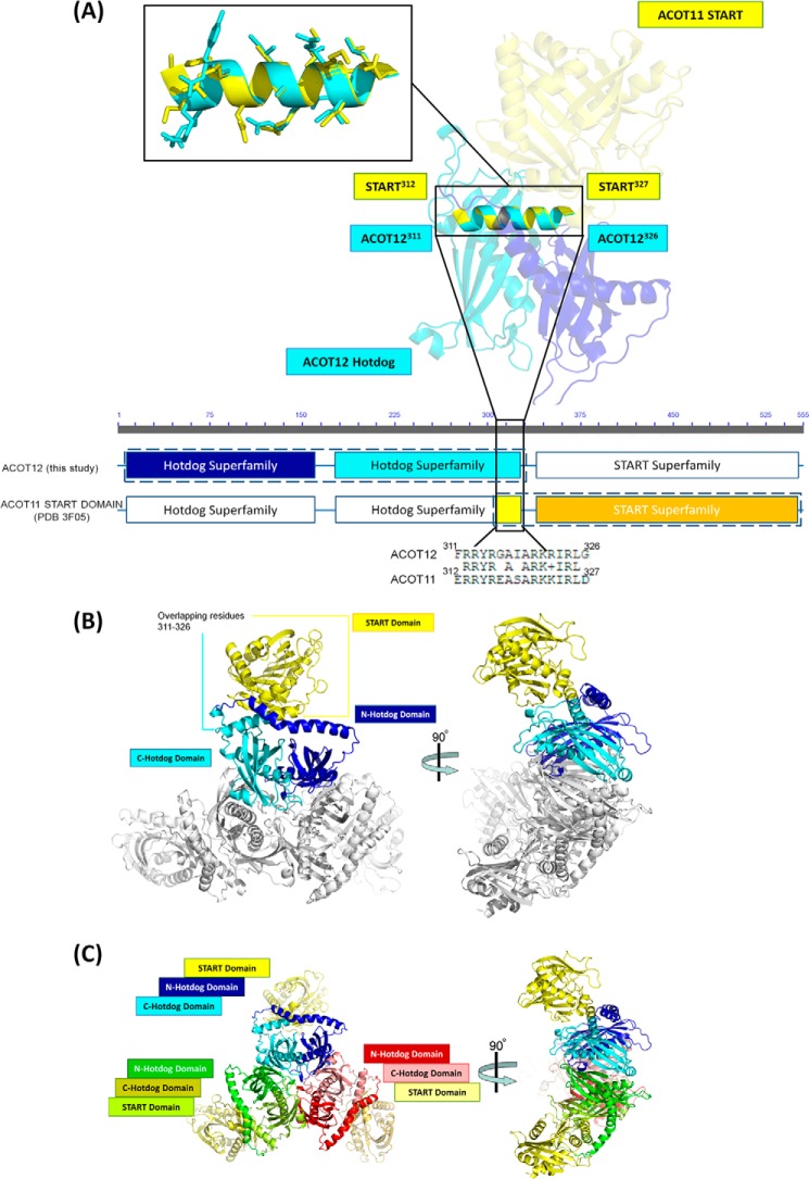
Acetyl-CoA plays a fundamental role in cell signaling and metabolic pathways, with its cellular levels tightly controlled through reciprocal regulation of enzymes that mediate its synthesis and catabolism. ACOT12, the primary acetyl-CoA thioesterase in the liver of human, mouse, and rat, is responsible for cleavage of the thioester bond within acetyl-CoA, producing acetate and coenzyme A for a range of cellular processes. The enzyme is regulated by ADP and ATP, which is believed to be mediated through the ligand-induced oligomerization of the thioesterase domains, whereby ATP induces active dimers and tetramers, whereas apo- and ADP-bound ACOT12 are monomeric and inactive. Here, using a range of structural and biophysical techniques, it is demonstrated that ACOT12 is a trimer rather than a tetramer and that neither ADP nor ATP exert their regulatory effects by altering the oligomeric status of the enzyme. Rather, the binding site and mechanism of ADP regulation have been determined to occur through two novel regulatory regions, one involving a large loop that links the thioesterase domains (Phe(154)-Thr(178)), defined here as RegLoop1, and a second region involving the C terminus of thioesterase domain 2 (Gln(304)-Gly(326)), designated RegLoop2. Mutagenesis confirmed that Arg(312) and Arg(313) are crucial for this mode of regulation, and novel interactions with the START domain are presented together with insights into domain swapping within eukaryotic thioesterases for substrate recognition. In summary, these experiments provide the first structural insights into the regulation of this enzyme family, revealing an alternate hypothesis likely to be conserved throughout evolution.

Keywords: ACOT12; ADP; ATP; Acetyl Coenzyme A (Acetyl-CoA); Allosteric Regulation; Metabolism; START Domain; Thioesterase.


Figures

FIGURE 1.
FIGURE 1.
Structure of ACOT12. A, domain architecture of ACOT12 comprised of two hotdog domains and C-terminal START domain. B, structure of the ACOT12 double hotdog domain protomer shown in schematic representation, with each hotdog domain in dark and light shades of the same color for each protomer. A 90° rotation highlights the presence of a β-bulge induced at each dimer interface. C, structure of the ACOT12 quaternary arrangement comprised of a trimer of hotdog dimers. D, structures of thioesterases containing the same hexameric arrangement built from either three double hotdog dimers (eukaryotes) or six single hotdog monomers (prokaryotes). Shown are structures of the thioesterase domains from B. halodurans (PDB code 1VPM (21)) a single hotdog fold thioesterase that forms a hexamer; the full length structure of ACOT7 (PDB codes 2Q2B and 2V1O (34)) and the structure of the double hotdog thioesterase from A. tumefaciens (PDB code 2GVH), which forms a back-to-back protomer.
FIGURE 2.
FIGURE 2.
The CoA binding motif of ACOT12. A, the two potential binding sites for CoA on ACOT12, with binding site I occupied by CoA as demonstrated by density at both 2 and 5σ of FoFc annealed omit maps and, in binding site II, a clear lack of density. B, the binding sites for CoA involving four separate binding regions; R1a (TAS55), R1b (STS84), R1c (Arg144), and R1d (KRFG237) for binding site I, whereas the binding site II contains steric clashes inhibiting CoA from occupying this site. C, location of CoA binding and active site residues on ACOT12 and ACOT7, demonstrating domain swapping between these two thioesterases for these critical functional regions. D, surface representation of ACOT12 with CoA shown as sticks and the substrate-binding tunnel is shown as spheres determined using Caver (38).
FIGURE 3.
FIGURE 3.
SAXS and SEC data confirm the quaternary structure observed from crystallographic data. A, ACOT12 scattering data (red dots) superimposed on the theoretical scattering curves (blue line) for an ACOT12 monomer, dimer, trimer, and tetramer using CRYSOL with χ values of 62, 25, 1.3, and 14.4, respectively. B, ACOT12-ADP scattering data with the theoretical scattering curves for ACOT12-ADP monomer, dimer, trimer, and tetramer superimposed with χ values of 25, 7, 1.8, and 5.5. C, SEC elution profiles for apo-ACOT12, ACOT12-ADP, and ACOT12-ATP plotted against known molecular weight standards to assess the native molecular weight demonstrate that neither ADP nor ATP disrupt oligomerization, consistent with the SAXS and crystallographic data.
FIGURE 4.
FIGURE 4.
Structure of ACOT12-ADP. A, the flexible regions 154–178 and 304–326 are visible in the ACOT12:ADP structure, revealing a 50° kink in the helix joining the two hotdog domains (loop I) at His150, causing residues 151–179 to link over the C-terminal α-helix. B, the hexamer arrangement of the ACOT12-ADP demonstrates its similarity to apo-ACOT12.
FIGURE 5.
FIGURE 5.
ACOT12 binding site of ADP. A, two potential binding sites, with binding site I having clear density at both 2 and 5σ in simulated annealed omit maps and binding site II exhibiting negligible density corresponding to ADP. B, residues involved in binding ADP at the two potential binding sites, with binding site I utilizing Arg312 and Arg313 to interact with the phosphate of ADP, whereas at binding site II, ADP is inhibited from binding due to the presence of Ser88 (equivalent to Gly in binding site I). C, superposition of ATP on the ADP site reveals the γ-phosphate of ATP clashes with Arg312 preventing the C-terminal helix from being locked into place. D, sequence alignment of human, mouse, and rat ACOT12 and ACOT11. The ADP and START domain binding residues are highlighted in blue and yellow, respectively.
FIGURE 6.
FIGURE 6.
ACOT12 activity and regulation activity. A, the ACOT12 thioesterase activity against acetyl-CoA concentration was plotted in GraphPad Prism using Michaelis-Menten kinetics (left panel). The activity of ACOT12 at 100 μm acetyl-CoA is enhanced by the presence of ATP and decreased by ADP, but this regulatory action is abolished in the Arg312/313 mutant, for which ATP and ADP (or GTP or GDP) show no significant differences (bottom panels, and summarized in right panel). The results shown in the bottom panel are from a single typical experiment repeated two or more times, with pooled data shown in the right panel representing the mean of activity (±) S.D. B, activity curves investigating the activity of ACOT12 and the Arg312/313 mutant over a range of ATP and ADP concentrations. Using the log concentrations of regulator the IC50 = 0.07290 mm for ADP and EC50 = 0.00297 mm for ATP were determined using Graphpad Prism.
FIGURE 7.
FIGURE 7.
The ACOT12 full-length model. A, structural alignment and superposition of ACOT12 with ACOT11 through overlapping residues 311–326. B, the ACOT11 START domain, shown in yellow, superimposed onto the ACOT12-ADP structure using the overlapping residues 311–326. C, the hexameric model of full-length ACOT12 containing all three START domains. Superimpositions were performed using Coot (26).

References

    1. Bartlett K., Eaton S. (2004) Mitochondrial β-oxidation. Eur. J. Biochem. 271, 462–469 - PubMed
    1. Wieland O., Weiss L., Eger-Neufeldt I. (1964) Enzymatic regulation of liver acetyl-CoA metabolism in relation to ketogenesis. Adv. Enzyme Regul. 2, 85–99 - PubMed
    1. Williamson D. H., Bates M. W., Krebs H. A. (1968) Activity and intracellular distribution of enzymes of ketone-body metabolism in rat liver. Biochem. J. 108, 353–361 - PMC - PubMed
    1. Wakil S. J., Abu-Elheiga L. A. (2009) Fatty acid metabolism: target for metabolic syndrome. J. Lipid Res. 50, S138–S143 - PMC - PubMed
    1. Brown M. S., Goldstein J. L. (1980) Multivalent feedback regulation of HMG CoA reductase, a control mechanism coordinating isoprenoid synthesis and cell growth. J. Lipid Res. 21, 505–517 - PubMed

LinkOut - more resources