. 2014 Aug 22;289(34):23745-52.
doi: 10.1074/jbc.M114.572149. Epub 2014 Jul 7.

Phosphatase 1 nuclear targeting subunit is an essential regulator of M-phase entry, maintenance, and exit

Affiliations

Phosphatase 1 nuclear targeting subunit is an essential regulator of M-phase entry, maintenance, and exit

Laura A Fisher et al. J Biol Chem. .

Abstract

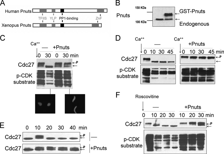
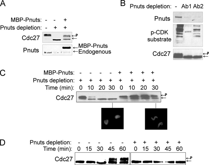
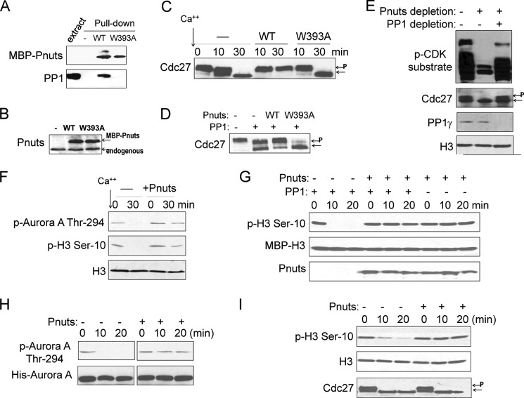
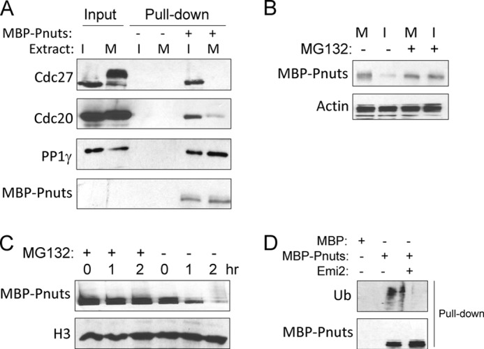
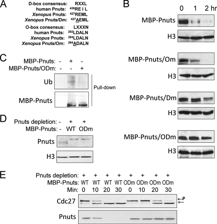
Mitotic progression is regulated largely through dynamic and reversible protein phosphorylation that is modulated by opposing actions of protein kinases and phosphatases. In this study, we show that phosphatase 1 nuclear targeting subunit (Pnuts) functions as a master regulator of mitosis by modulating protein phosphatase 1 (PP1). Overexpression of Pnuts in Xenopus egg extracts inhibited both mitotic and meiotic exit. Immunodepletion of Pnuts from egg extracts revealed its essential functions in mitotic entry and maintenance. The level of Pnuts oscillates during the cell cycle and peaks in mitosis. Pnuts destruction during M-phase exit is mediated by the anaphase-promoting complex/cyclosome (APC/C)-targeted ubiquitination and proteolysis, and conserved destruction motifs of Pnuts. Disruption of Pnuts degradation delayed M-phase exit, suggesting it as an important mechanism to permit M-phase exit.

Keywords: APC/C; Cell Cycle; Mitosis; Phosphatase; Phosphoprotein Phosphatase 1 (PP1); Pnuts; Xenopus.


Figures

FIGURE 1.
FIGURE 1.
Pnuts regulates M-phase exit. A, schematic representation of human and Xenopus Pnuts proteins showing domains conserved from Xenopus to human. TFIIS, transcription elongation factor II-like domain; YLP, telomeric repeat binding factor 2-binding motif; ZnF, zinc finger domain. B, the addition of purified recombinant Xenopus Pnuts in Xenopus egg extracts. The relative amount of endogenous and exogenous Pnuts is shown by immunoblotting using an anti-Pnuts antibody. C, calcium (40 nm) was added to CSF Xenopus egg extracts with or without exogenous Pnuts as in panel B. Samples were taken at the indicated time points and immunoblotted for Cdc27 and Phospho-CDK (p-CDK) substrates. Phosphorylated Cdc27 is indicated by P. Extracts were supplemented with sperm nuclei and monitored for the morphology of sperm nuclei, stained with DAPI. D, calcium was added to CSF Xenopus egg extracts with or without exogenous Pnuts to induce M-phase exit. Samples were taken at the indicated time points and immunoblotted for Cdc27 and Phospho-CDK substrates. E, cycling extracts in the absence or presence of exogenous Pnuts were examined for Cdc27 phosphorylation. F, the CDK inhibitor roscovitine (0.5 mm) was added to CSF extracts in the absence or presence of exogenous Pnuts and incubated at room temperature for 30 min. Extract samples were taken at the indicated time points and immunoblotted for Cdc27 and phospho-CDK substrates.
FIGURE 2.
FIGURE 2.
Pnuts is an essential M-phase regulator. A, Pnuts was immunodepleted from CSF extracts as described under “Experimental Procedures.” Recombinant Pnuts was used to restore Pnuts expression in the depleted extract. Extracts were incubated at room temperature for 30 min and immunoblotted for Pnuts and Cdc27. Phosphorylated Cdc27 is indicated by P. B, Pnuts was immunodepleted from CSF extracts using two antibodies (Ab1 and Ab2). Extracts were incubated at room temperature for 30 min and immunoblotted for Pnuts, Cdc27, and phospho-CDK (p-CDK) substrates. C, as in panel A, Pnuts was immunodepleted from CSF extracts with or without adding back MBP-Pnuts. Extract samples were harvested at the indicated time points and analyzed by immunoblotting and the morphology of sperm nuclei. D, cycling extracts were mock-treated (−), or immunodepleted of Pnuts (+), and immunoblotted for Cdc27.
FIGURE 3.
FIGURE 3.
Pnuts regulates M-phase through PP1. A, wild-type, but not W393A mutant, Pnuts binds PP1. MBP-Pnuts bound to amylose resin was incubated in Xenopus egg extracts and then reisolated as described under “Experimental Procedures.” The input extract and pulldown products were examined by immunoblotting for MBP and PP1. B, supplementation of WT or W393A MBP-Pnuts in Xenopus egg extracts. Extract samples were immunoblotted using anti-Pnuts antibody. C, as in Fig. 1C, calcium was added into CSF extracts to induce M-phase exit, with or without WT or W393A Pnuts. Extract samples were harvested after 30 min of incubation and analyzed by immunoblotting. Phosphorylated Cdc27 is indicated by P. D, PP1 (New England Biolabs, 0.42 unit/μl) was added into CSF extracts to induce M-phase exit, with or without WT or W393A Pnuts. Extract samples were harvested after 30 min of incubation and analyzed by immunoblotting. E, CSF extracts were mock-treated (−), depleted of Pnuts as in Fig. 2A, or co-depleted of Pnuts and PP1γ, and incubated at room temperature for 30 min. Immunoblotting of phospho-CDK (p-CDK) substrates, Cdc27, and PP1 is shown. F, CSF extracts with or without supplementation of MBP-Pnuts were treated with calcium. Extract samples were collected at the indicated time points and analyzed by immunoblotting for phospho-Aurora A (p-Aurora A), phospho-H3 (p-H3), and H3. G, the MBP-H3-S10 substrate was generated and prephosphorylated as described under “Experimental Procedures.” PP1 was used to dephosphorylate the substrate in vitro, with or without the addition of Pnuts protein. Immunoblots of phopho-H3 Ser-10, MBP, and Pnuts are shown. H, purified WT Aurora A was added to interphase extracts with or without MBP-Pnuts and incubated at room temperature. Samples were taken at the indicated time points and immunoblotted using phospho-Aurora A and His tag antibodies. I, CSF extracts were diluted (1:5) in the PP1-containing buffer (New England Biolabs) with or without Pnuts. Samples were collected at the indicated time points and analyzed by immunoblotting using phospho-H3, H3, and Cdc27 antibodies.
FIGURE 4.
FIGURE 4.
Cell cycle-dependent regulation of Pnuts expression. A, immunoblotting of Pnuts in M-phase (M, CSF extract) or interphase (I, released from the CSF extract) in two independent sets of extracts. H3 and Aurora B are shown as loading controls. B, cycling extract samples were taken at the indicated time points and then analyzed by immunoblotting for Pnuts expression and Cdc27 phosphorylation during the cell cycle. Phosphorylated Cdc27 is indicated by P. C, MBP-Pnuts was added to M-phase extract along with calcium to induce M-phase exit, and extract samples were taken at the indicated time points and analyzed for Cdc27 and Pnuts. D, MBP-Pnuts was added to CSF extract (M-phase) or interphase extract (released from the same CSF extract) and incubated as indicated. Extracts were then analyzed by immunoblotting using MBP tag and H3 antibodies.
FIGURE 5.
FIGURE 5.
Regulation of Pnuts stability through APC/C-mediated ubiquitination and proteolysis. A, MBP-Pnuts (+) or control MBP (−) was reisolated from either interphase (I) or M-phase (M) extracts, as described under “Experimental Procedures,” and analyzed by immunoblotting. The input contains 10% of the extracts used. B, MBP-Pnuts was added into M-phase or interphase extracts and incubated for 1 h, with or without proteasome inhibitor MG132. Immunoblotting of MBP and actin is shown. C, MBP-Pnuts and MG132 were added into interphase extracts and incubated over time, as indicated. Extract samples were then analyzed by immunoblotting or MBP and H3. D, ubiquitination (Ub) of Pnuts was measured by immunoblotting of MBP-Pnuts reisolated from extract with or without the APC/C inhibitor Emi2, using anti-ubiquitin and anti-MBP antibodies.
FIGURE 6.
FIGURE 6.
Pnuts stability is regulated through conserved destruction box motifs. A, alignment of the D- and O-box sequences of human and Xenopus Pnuts and the mutations made to them in this study. B, Pnuts was mutated in the O-box (Om), D-box (Dm), or both boxes (ODm), as in panel A. The resulting mutants, along with WT Pnuts, were added into interphase extracts, incubated over time, and measured by immunoblotting for their stability. C, as in Fig. 5D, ubiquitination (Ub) of WT or ODm Pnuts was measured by immunoblotting. D and E, endogenous Pnuts was immunodepleted from CSF extracts, which were then supplemented with either WT or ODm Pnuts. The extracts were then treated with Ca2+, collected at the indicated time points, and analyzed by immunoblotting for Cdc27 and Pnuts. Phosphorylated Cdc27 is indicated by P.

References

    1. Janssen A., Medema R. H. (2011) Mitosis as an anti-cancer target. Oncogene 30, 2799–2809 - PubMed
    1. Nigg E. A. (2001) Mitotic kinases as regulators of cell division and its checkpoints. Nat. Rev. Mol. Cell Biol. 2, 21–32 - PubMed
    1. Malumbres M., Harlow E., Hunt T., Hunter T., Lahti J. M., Manning G., Morgan D. O., Tsai L. H., Wolgemuth D. J. (2009) Cyclin-dependent kinases: a family portrait. Nat. Cell Biol. 11, 1275–1276 - PMC - PubMed
    1. Lens S. M., Voest E. E., Medema R. H. (2010) Shared and separate functions of Polo-like kinases and Aurora kinases in cancer. Nat. Rev. Cancer 10, 825–841 - PubMed
    1. Mochida S., Hunt T. (2012) Protein phosphatases and their regulation in the control of mitosis. EMBO Rep. 13, 197–203 - PMC - PubMed

Publication types

LinkOut - more resources