

Histological-subtypes and anatomical location correlated in meningeal brain tumors (meningiomas)
- PMID: 25002762
- PMCID: PMC4078607
- DOI: 10.4103/0976-3147.133568
Histological-subtypes and anatomical location correlated in meningeal brain tumors (meningiomas)
Abstract
Context: Not enough literature is available to suggest a link between the histological subtypes of intracranial meningeal brain tumors, called 'meningiomas' and their location of origin.
Aim: The evidence of correlation between the anatomical location of the intracranial meningiomas and the histopathological grades will facilitate specific diagnosis and accurate treatment.
Materials and methods: The retrospective study was conducted in a single high-patient-inflow Neurosurgical Center, under a standard and uniform medical protocol, over a period of 30 years from December 1982 to December 2012. The records of all the operated 729 meningiomas were analyzed from the patient files in the Medical Records Department. The biodata, x-rays, angiography, computed tomography (CT) scans, imaging, histopathological reports, and mortality were evaluated and results drawn.
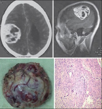
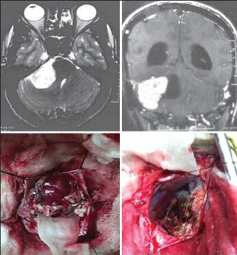
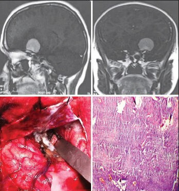
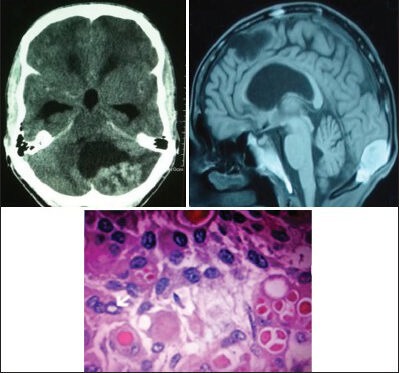
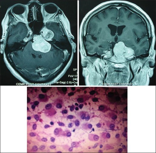
Results: The uncommon histopathological types of meningiomas (16.88%) had common locations of origin in the sphenoid ridge, posterior parafalcine, jugular foramen, peritorcular and intraventricular regions, cerebellopontine angle, and tentorial and petroclival areas. The histopathological World Health Organization (WHO) Grade I (Benign Type) meningiomas were noted in 89.30%, WHO Grade II (Atypical Type) in 5.90%, and WHO Grade III (Malignant Type) in 4.80% of all meningiomas. Meningiomas of 64.60% were found in females, 47.32% were in the age group of 41-50 years, and 3.43% meningiomas were found in children. An overall mortality of 6.04% was noted. WHO Grade III (malignant meningiomas) carried a high mortality (25.71%) and the most common sites of meningiomas with high mortality were: The cerebellopontine angles, intraventricular region, sphenoid ridge, tuberculum sellae, and the posterior parafalcine areas.
Conclusion: The correlation between the histological subtypes and the anatomical location of intracranial meningeal brain tumors, called meningiomas, is evident, but further research is required to establish the link.
Keywords: Anatomical origin; correlation; histological types; intracranial meningiomas.
Conflict of interest statement
Figures
References
-
- Okonkwo DO, Laws ER. Meningiomas: Historical perspective. In: Lee JH, editor. Meningiomas: Diagnosis, Treatment, and Outcome. London: Springer-Verlag; 2009. pp. 3–11.
-
- Perry A, Stafford SL, Scheithauer BW, Suman VJ, Lohse CM. Meningioma grading: An analysis of histologic parameters. Am J Surg Pathol. 1997;21:1455–65. - PubMed
-
- Kaye H. Posterior fossa meningiomas. In: Sindou M, editor. Practical Handbook of Neurosurgery: From Leading Neurosurgeons. Vol. 2. Germany: Springer-Verlag; 2009. p. 181.
-
- Bondy M, Ligon BL. Epidemiology and etiology of intracranial meningiomas: A review. J Neurooncol. 1996;29:197–205. - PubMed
-
- Marin Sanabria EA, Ehara K, Tamaki N. Surgical experience with skull base approaches for foramen magnum meningioma. Neurol Med Chir (Tokyo) 2002;42:472–80. - PubMed
LinkOut - more resources
Full Text Sources
Other Literature Sources