. 2014:2014:940186.
doi: 10.1155/2014/940186. Epub 2014 Jun 5.

The Ginkgo biloba Extract EGb 761 Modulates Proteasome Activity and Polyglutamine Protein Aggregation

Affiliations

The Ginkgo biloba Extract EGb 761 Modulates Proteasome Activity and Polyglutamine Protein Aggregation

Marcel Stark et al. Evid Based Complement Alternat Med. 2014.

Abstract

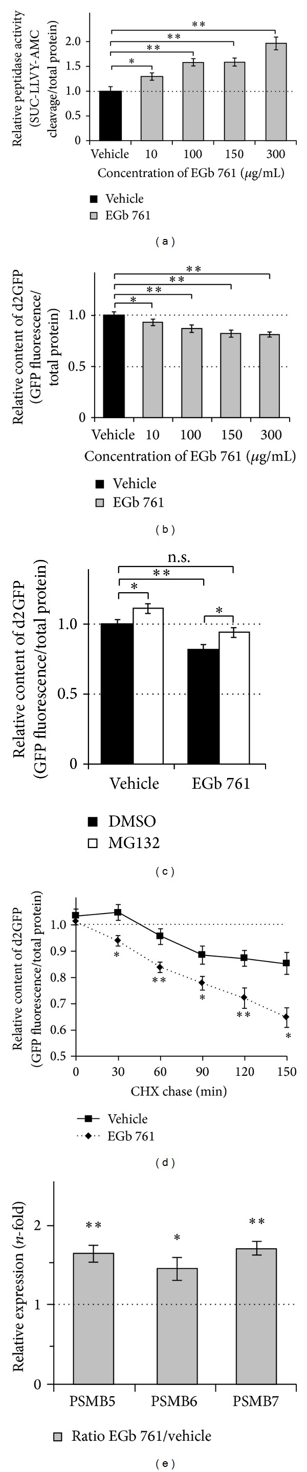
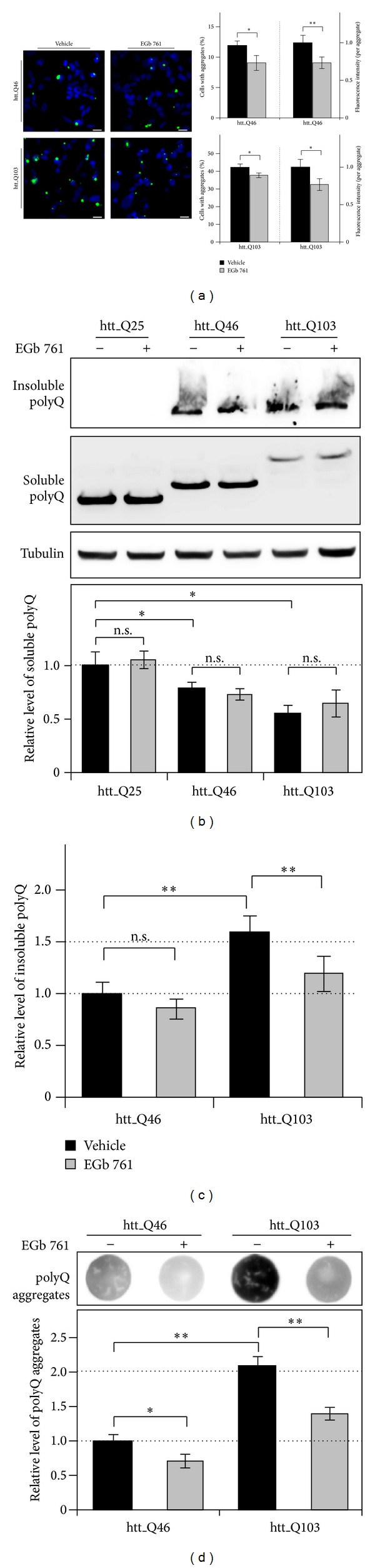
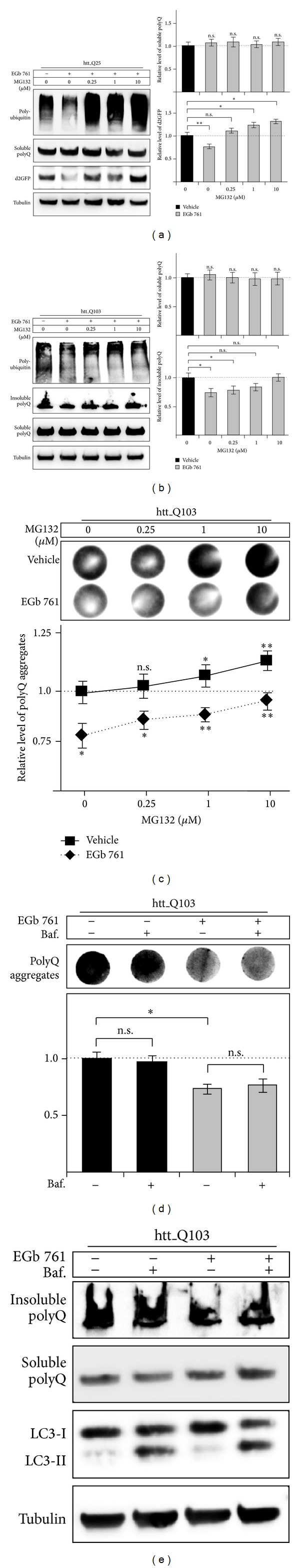
The standardized Ginkgo biloba extract EGb 761 has well-described antioxidative activities and effects on different cytoprotective signaling pathways. Consequently, a potential use of EGb 761 in neurodegenerative diseases has been proposed. A common characteristic feature of a variety of such disorders is the pathologic formation of protein aggregates, suggesting a crucial role for protein homeostasis. In this study, we show that EGb 761 increased the catalytic activity of the proteasome and enhanced protein degradation in cultured cells. We further investigated this effect in a cellular model of Huntington's disease (HD) by employing cells expressing pathologic variants of a polyglutamine protein (polyQ protein). We show that EGb 761 affected these cells by (i) increasing proteasome activity and (ii) inducing a more efficient degradation of aggregation-prone proteins. These results demonstrate a novel activity of EGb 761 on protein aggregates by enhancing proteasomal protein degradation, suggesting a therapeutic use in neurodegenerative disorders with a disturbed protein homeostasis.


Figures

Figure 1
Figure 1
Effects of EGb 761 on basal proteasome activity. (a) HEK293 cells were treated for 24 h with indicated concentrations of EGb 761. Analysis of proteasomal peptidase (chymotrypsin-like) activity was assessed by the hydrolysis of SUC-LLVY-AMC in total cell lysates. Fluorescence of the cleaved AMC moiety was measured in the presence or absence of MG132 to achieve peptidase specificity. Values were adjusted to total protein content. Activity of vehicle-treated cells was arbitrarily set to 1; n = 4. (b–d) HEK293 cells with stable expressions of the proteasome reporter protein d2GFP (d2GFP-HEK) were treated for 24 h with indicated concentrations of EGb 761. Measurement of GFP fluorescence was used to investigate proteasomal degradation of d2GFP proteins. All achieved fluorescence intensities were finally adjusted to total protein content. (b) Cells were incubated with increasing concentrations of EGb 761 to investigate the specific, dose-depending effect on proteasome activity (a). Measurement of GFP fluorescence was used to assess the remaining d2GFP protein content as an indicator for an enhanced protein degradation by the proteasome. Values of vehicle-treated cells were arbitrarily set to 1. n = 5. (c) Previous assayed cells ((b); vehicle and 150 μg/mL EGb 761 treatments) were additionally incubated for 2 h with the proteasome inhibitor MG132 or DMSO as control. The addition of MG132 led to an increase of fluorescence intensities in control and EGb 761-treated cells. Inhibition of proteasome activity showed the specific modulation of GFP fluorescence through proteasomal d2GFP degradation. Values of vehicle-treated cells without MG132 were arbitrarily set to 1. n = 4. (d) Cells were treated for 24 h with 150 μg/mL EGb 761 or vehicle, followed by a chase with cycloheximide (CHX) to block synthesis of new d2GFP. Degradation kinetics of d2GFP was analyzed by measuring GFP fluorescence every 30 min. Fluorescence decay induced by CHX indicated the specificity of proteasomal d2GFP degradation. Values of each treatment at zero minutes were arbitrarily set to 1; n = 3. (e) HEK293 cells were treated for 2 h with 150 μg/mL EGb 761 or vehicle and RNA was extracted for qRT-PCR analysis. Relative expression ratio of proteasome genes PSMB5, PSMB6, and PSMB7 in EGb 761-treated cells to vehicle-treated cells is shown; n = 3. (a–e) All values are reported as mean ± S.D. *P < 0.05 and **P < 0.01.
Figure 2
Figure 2
Modulation of proteasome activity by EGb 761 in cells expressing polyQ proteins. (a, c-d) HEK293 or (b) d2GFP-HEK cells were transiently transfected to express the polyQ fusion proteins with glutamine expansions of 25 (htt_Q25), 46 (htt_Q46), and 103 repeats (htt_Q103). Subsequent to settlement for 24 h cells were treated with 150 μg/mL EGb 761 or vehicle for different time periods and further investigated. (a) HEK293 cells expressing eGFP and polyQ proteins (htt_Q25, Q46, and Q103) were treated for 24 h with EGb 761 or vehicle, followed by analysis of proteasomal peptidase activity in total cell lysates. Fluorescence of AMC moiety resulting from peptidase hydrolysis of SUC-LLVY-AMC was measured in the presence or absence of MG132 to achieve peptidase specificity. Values were adjusted to total protein content. Activity of vehicle-treated eGFP-expressing cells was arbitrarily set to 1; n = 3. (b) d2GFP-HEK cells expressing polyQ proteins (htt_Q25, Q46, and Q103) were treated for 24 h with EGb 761 or vehicle. Cell lysates of each sample were collected and analyzed by immunoblotting. Protein levels of d2GFP and proteasome subunit α1 were analyzed with their corresponding antibody to assess proteasome activity and status. Densitometric values of vehicle-treated and htt_Q25-expressing cells were arbitrarily set to 1; n = 4. (c-d) HEK293 cells expressing htt_Q25 and htt_Q103 for 24 h were analyzed by qRT-PCR for transcript levels of PSMB5, PSMB6, and PSMB7. (c) Quantification of basal expression ratio in cells expressing htt_Q103 to htt_Q25 (without EGb 761 treatment). (d) Analysis of cells additionally treated with EGb 761 or vehicle for 2 h. Expression ratio of proteasome genes in cells treated with EGb 761 compared to vehicle; n = 3. (a–d) All values are reported as mean ± S.D. *P < 0.05 and **P < 0.01.
Figure 3
Figure 3
Modulating effects of EGb 761 on aggregation of polyQ proteins. (a–d) HEK293 cells expressing htt_Q46, htt_Q103, and (b) htt_Q25 were treated for 48 h with 150 μg/mL EGb 761 or vehicle. (a) Fluorescence microscopy revealed significantly more polyQ aggregates in cells expressing htt_Q103 compared to htt_Q46. Note that most aggregates are formed as nuclear inclusions. Percentage of cells exhibiting fluorescent aggregates (%) was determined by plotting total amount of polyQ aggregates against total DAPI positive cells. Total cell and aggregate amount resulted from at least three independent experiments (more than 100 cells counted). Relative fluorescence intensity (a.u.) of aggregates was quantified by calculating aggregate mean intensity with aggregate area (pixel∗au). Representative pictures are shown. Scale bar: 20 μm. (b) Cells expressing htt_Q25, htt_Q46, and htt_Q103 with EGb 761 or vehicle treatments were analyzed by immunoblotting. PolyQ proteins were detected with anti-eGFP antibody showing SDS-soluble protein bands. Cells expressing htt_Q46 and htt_Q103 showed aggregated SDS-insoluble proteins in the stacking gel (different exposure times were used). Densitometric values of soluble polyQ proteins from vehicle-treated htt_Q25-expressing cells were arbitrarily set to 1; n = 4. (c) Analysis of polyQ aggregation by using densitometric data of insoluble polyQ proteins from previous immunoblots (b). Values of vehicle-treated htt_Q46-expressing cells were arbitrarily set to 1; n = 4. (d) Whole cell extracts from cells expressing htt_Q46 and htt_Q103 with EGb 761 or vehicle treatment (samples from d) were subjected to a filter retardation assay. Detection of polyQ proteins trapped on nitrocellulose membrane revealed highly aggregated polyQ proteins in htt_Q103 compared to htt_Q46. Densitometric values of polyQ aggregates from vehicle-treated htt_Q46-expressing cells were arbitrarily set to 1; n = 4. (a–d) All values are reported as mean ± S.D. *P < 0.05 and **P < 0.01.
Figure 4
Figure 4
Effects of EGb 761 on degradation of polyQ proteins and polyQ aggregation. (a) d2GFP-HEK cells expressing htt_Q25 and (b–d) HEK293 cells expressing htt_Q103 were treated with EGb 761 or vehicle and subsequently chased with proteasome (MG132) or autophagy inhibitor (bafilomycin) to assess polyQ degradation and aggregation. (a) d2GFP-HEK cells expressing htt_Q25 were treated for 48 h with 150 μg/mL EGb 761 or vehicle. Then, cells were incubated for 4 h with or without increasing concentrations of MG132. Protein levels of whole cell extracts were analyzed by immunoblotting to their corresponding antibodies. Protein levels of short-lived, unstable proteins (polyubiquitin, d2GFP) accumulated with proteasome inhibition while levels of long-lived polyQ proteins were not significantly altered. Values of d2GFP or polyQ protein of vehicle-treated cells without MG132 were arbitrarily set to 1; n = 4. (b-c) HEK293 cells expressing htt_Q103 were treated for 48 h with 150 μg/mL EGb 761 or vehicle. Then, cells were subsequently incubated for 4 h with or without increasing concentrations of MG132. (b) Whole cell extracts were subjected to a filter retardation assay to assess polyQ aggregates, induced by pharmacologic proteasome inhibition. For the detection of aggregates of polyQ proteins trapped on nitrocellulose membrane an anti-eGFP antibody was used. Densitometric values of vehicle-treated cells without MG132 were arbitrarily set to 1; n = 5. (c) Protein levels of whole cell extracts (samples from b) were analyzed by immunoblotting to their corresponding antibodies. Protein levels of unstable, misfolded polyQ proteins accumulated with proteasome inhibition while levels of stable, soluble polyQ proteins were not altered. Values of soluble or insoluble polyQ proteins of vehicle-treated cells without MG132 were arbitrarily set to 1; n = 3. (d) HEK293 cells expressing htt_Q103 were treated for 48 h with 150 μg/mL EGb 761 or vehicle. Then, cells were subsequently incubated for 3 h with or without 1 μM of the lysosomal inhibitor bafilomycin A1 (denoted bafi.) to investigate the autophagic flux. Whole cell extracts were analyzed by immunoblotting or filter retardation assay. Cells treated with EGb 761 showed no significant alteration with bafilomycin treatment in protein levels of LC3-I to LC3-II, soluble and insoluble polyQ, indicating no direct effect on autophagy by EGb 761. Immunoblotting and filter retardation assay confirmed significant changes in aggregated polyQ proteins by EGb 761 from previous experiments (Figures 3(b)–3(d) and Figures 4(b)-4(c)). Densitometric values of vehicle-treated cells without bafilomycin were arbitrarily set to 1; n = 3. (a–d) All values are reported as mean ± S.D. *P < 0.05 and **P < 0.01.

Similar articles

Cited by

References

    1. van Beek TA. Chemical analysis of Ginkgo biloba leaves and extracts. Journal of Chromatography A. 2002;967(1):21–55. - PubMed
    1. Mahadevan S, Park Y. Multifaceted therapeutic benefits of Ginkgo biloba L.: chemistry, efficacy, safety, and uses. Journal of Food Science. 2008;73(1):R14–R19. - PubMed
    1. Chan P-C, Xia Q, Fu PP. Ginkgo biloba leave extract: biological, medicinal, and toxicological effects. Journal of Environmental Science and Health C: Environmental Carcinogenesis & Ecotoxicology Reviews. 2007;25:211–244. - PubMed
    1. Janßen IM, Sturtz S, Skipka G, Zentner A, Garrido MV, Busse R. Ginkgo biloba in Alzheimer’s disease: a systematic review. Wiener Medizinische Wochenschrift. 2010;160(21-22):539–546. - PubMed
    1. Horáková L, Licht A, Sandig G, Jakstadt M, Duracková Z, Grune T. Standardized extracts of flavonoids increase the viability of PC12 cells treated with hydrogen peroxide: effects on oxidative injury. Archives of Toxicology. 2003;77(1):22–29. - PubMed

LinkOut - more resources