

Spinal drop metastasis in myxopapillary ependymoma: a case report and a review of treatment options
- PMID: 25002955
- PMCID: PMC4083675
- DOI: 10.4081/rt.2014.5404
Spinal drop metastasis in myxopapillary ependymoma: a case report and a review of treatment options
Abstract
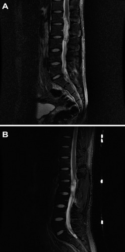
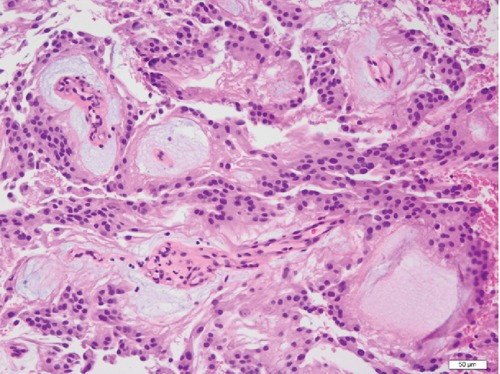
Myxopapillary ependymoma (MPE) is a World Health Organization grade I ependymoma that is quite rare and generally thought to be benign. Possible drop metastasis from MPE has been reported three times in the literature; in each case there were cotemporaneous additional MPE lesions. We report the case of a man who had a piecemeal gross total resection of a MPE at L1-L3 followed by adjuvant external beam radiotherapy (EBRT) who presented sixteen months later with a lesion in the thecal sac consistent with drop metastasis. A subtotal resection and adjuvant EBRT were performed. The patient has been disease-free in follow-up 27 months from the second surgery. A review of the literature regarding the treatment for MPE showed that gross total resection is optimal initial management. Several retrospective studies supported the role of adjuvant radiotherapy in enhancing local control and progression-free survival. Chemotherapy has a minimal role in the management of MPE.
Keywords: adjuvant radiotherapy; drop metastasis; ependymoma; myxopapillary.
Conflict of interest statement
Conflict of Interests: the authors declare no potential conflict of interests.
Figures
Similar articles
-
Spinal myxopapillary ependymoma with interval drop metastasis presenting as cauda equina syndrome: case report and review of literature.J Spine Surg. 2016 Sep;2(3):216-221. doi: 10.21037/jss.2016.08.06. J Spine Surg. 2016. PMID: 27757435 Free PMC article.
-
Long-Term Surgical Resection Outcomes of Pediatric Myxopapillary Ependymoma: Experience of Two Centers and Brief Literature Review.World Neurosurg. 2020 Apr;136:e245-e261. doi: 10.1016/j.wneu.2019.12.128. Epub 2019 Dec 30. World Neurosurg. 2020. PMID: 31899399 Review.
-
Outcomes following myxopapillary ependymoma resection: the importance of capsule integrity.Neurosurg Focus. 2015 Aug;39(2):E8. doi: 10.3171/2015.5.FOCUS15164. Neurosurg Focus. 2015. PMID: 26235025
-
Clinical management and prognosis of spinal myxopapillary ependymoma: a single-institution cohort of 72 patients.Eur Spine J. 2023 Jul;32(7):2459-2467. doi: 10.1007/s00586-023-07690-9. Epub 2023 Apr 7. Eur Spine J. 2023. PMID: 37027035
-
Primary Seeding of Myxopapillary Ependymoma: Different Disease in Adult Population? Case Report and Review of Literature.World Neurosurg. 2017 Mar;99:812.e21-812.e26. doi: 10.1016/j.wneu.2016.12.022. Epub 2016 Dec 29. World Neurosurg. 2017. PMID: 28040529 Review.
Cited by
-
A rare case of an intramedullary metastasis of a myxopapillary ependymoma.Surg Neurol Int. 2019 May 10;10:83. doi: 10.25259/SNI-96-2019. eCollection 2019. Surg Neurol Int. 2019. PMID: 31528421 Free PMC article.
-
Multifocal lumbar myxopapillary ependymoma presenting with drop metastasis: a case report and review of the literature.Spinal Cord Ser Cases. 2022 Apr 22;8(1):43. doi: 10.1038/s41394-022-00513-x. Spinal Cord Ser Cases. 2022. PMID: 35459220 Free PMC article. Review.
-
Sacral ependymoma presents 20 years after initial posterior fossa lesion.BMJ Case Rep. 2023 Oct 19;16(10):e256611. doi: 10.1136/bcr-2023-256611. BMJ Case Rep. 2023. PMID: 37857539 Free PMC article.
-
Outcomes and Pattern of Care for Spinal Myxopapillary Ependymoma in the Modern Era-A Population-Based Observational Study.Cancers (Basel). 2024 May 25;16(11):2013. doi: 10.3390/cancers16112013. Cancers (Basel). 2024. PMID: 38893133 Free PMC article.
-
Myxopapillary ependymoma: a SEER analysis of epidemiology and outcomes.J Neurooncol. 2016 Sep;129(2):251-8. doi: 10.1007/s11060-016-2167-0. Epub 2016 Jun 15. J Neurooncol. 2016. PMID: 27306443
References
-
- Reni M, Gatta G, Mazza E, Vecht C. Ependymoma. Crit Rev Oncol Hematol. 2007;63:81–9 - PubMed
-
- Davis C, Barnard RO. Malignant behavior of myxopapillary ependymoma. Report of three cases. J Neurosurg. 1985;62:925–9 - PubMed
-
- Fassett DR, Pingree J, Kestle JR. The high incidence of tumor dissemination in myxopapillary ependymoma in pediatric patients. Report of five cases and review of the literature. J Neurosurg. 2005;102:59–64 - PubMed
-
- Wang M, Wang H, Zhou Y, et al. Myxopapillary ependymoma in the third ventricle area and sacral canal: dropped or retrograde metastasis? Neurol Med Chir (Tokyo). 2013;53:237–41 - PubMed
Publication types
LinkOut - more resources
Full Text Sources
Other Literature Sources
